Honey, I Shrunk the Columns: Recent Advances in the Design and Use of Narrow Internal Diameter UHPLC Columns
The quest for increased sensitivity for low-concentration samples continues at an accelerated pace, especially in the pharmaceutical and clinical environments. High-end mass spectrometry instruments can enable the analyst to detect analytes at attomolar concentrations, but can be prohibitively expensive and require well-trained scientists to operate and interpret the data. Another approach to improve the sensitivity of high performance liquid chromatography (HPLC) assays is by decreasing the internal diameter (i.d.) of the column. Reducing the column’s internal diameter down from 4.6 mm to 2.1 mm has been in practice for several decades. However, extra caution is required when considering going down below 2.1-mm i.d. into the “capillary-flow” format. This paper describes advances in column design that enable the analyst to achieve high sensitivity and efficiency when reducing the internal diameter of the column. After an introduction outlining the theory of increased sensitivity with narrow and capillary-scale internal diameter columns, a novel 1.5-mm i.d. column format will be presented detailing how sensitivities comparable to capillary-like separations can be achieved with this column geometry. Application examples will demonstrate the performance benefits of utilizing a 1.5-mm i.d. column geometry.
High performance liquid chromatography (HPLC) assays play a crucial role in pharmaceutical and clinical laboratories by enabling the precise and accurate quantification of compounds in complex matrices. Sensitivity in HPLC assays is paramount as it directly impacts the ability to detect trace amounts of target analytes. This attribute is particularly important in pharmaceutical research and clinical diagnostics, where low concentrations of drugs or metabolites can have significant therapeutic or diagnostic implications. Enhanced sensitivity allows for better characterization of drug formulations, detection of impurities, and monitoring of drug levels in biological samples, thereby ensuring the safety, efficacy, and quality of pharmaceutical products and patient care.
In liquid chromatography (LC), there are several strategies to improve the sensitivity of a given assay. Mass spectrometry (MS) as the detector is one such approach to improve sensitivity and specificity. The choice of detector is critical to achieving the required sensitivity for the quantification of target analytes. Ultraviolet (UV) detectors are commonly used in HPLC because of their simplicity, affordability, and widespread applicability. However, they typically offer lower sensitivity compared to mass spectrometer (MS) detectors. MS detectors, such as electrospray ionization (ESI) or atmospheric-pressure chemical ionization (APCI), provide significantly higher sensitivity by ionizing analyte molecules and detecting them based on their mass-to-charge ratio (m/z). This attribute allows for the detection and quantification of analytes at much lower concentrations, making MS particularly valuable for the analysis of complex samples and trace-level components. While UV detectors are sufficient for many routine HPLC applications, MS detectors are indispensable for demanding analyses requiring high sensitivity and specificity, especially in pharmaceutical research and clinical diagnostics where trace-level detection is often essential for accurate and reliable results.
Another strategy for enhancing sensitivity is to decrease the internal diameter (i.d.) of the column. This practice has been used for several decades, first with scaling methods from 7.8-mm i.d. columns down to 4.6-mm i.d. columns. With the advent of ultrahigh-pressure liquid chromatography (UHPLC) instruments, methods could be scaled down even further, going from 4.6-mm i.d. columns down to 2.1-mm i.d. and even to 1.0-mm i.d. As methods are scaled down, the effects of extracolumn dispersion from the instrument become more pronounced, leading to a decrease in sensitivity even if a method is scaled correctly. With 1.0-mm i.d. columns, this effect becomes prominent, even with well plumbed instruments with minimal extracolumn volume.
The purpose of this paper is to describe a solution to this fundamental problem using a 1.5-mm i.d. column. After a review on the fundamentals of scaling methods down from larger internal diameter columns to smaller ones, an examination of the novel 1.5-mm i.d. column will be performed. Application examples will be used to demonstrate the performance benefits of using this column geometry for large molecule separations.
Fundamentals of Method Scaling
A transfer of HPLC methods to UHPLC requires scaling down from columns with larger internal diameter (for example, 4.6-mm i.d.), to columns with a smaller internal diameter (such as ~2.1-mm i.d.), and from long columns (for example, 15 cm length), to short columns (such as 5 cm length), in addition to the reduction of particle sizes (for example, from 5 μm to 2 μm). To ensure equivalent chromatographic separation, it is also necessary to scale the flow rate, injection volume and the gradient parameters.
Adjusting the Column Length
The first step is to determine the appropriate column length to maintain the same separation. Keeping the same column length while decreasing the particle size will increase the number of theoretical plates as well as back pressure. Therefore, when decreasing particle size, column length can be shortened without losing resolution (equation 1):

Column length (L), Flow Rate (f), Injection Volume (V), Time (t), Particle size (d)
Scaling the Flow Rate
Decreasing the internal diameter of the column (from 4.6 mm to 2.1 mm) requires recalculation of column flow rate to maintain linear velocity. Linear velocity is defined as the distance which mobile phase travels over time (cm/min), whereas flow rate is the volume of mobile phase that travels over time (mL/min). To maintain the same linear velocity through a column with a smaller internal diameter, the flow rate must be decreased proportionally to the column internal diameter according to equation 2 below:

Column length (L), Flow Rate (f), Injection Volume (V), Time (t), Particle size (d)
Scaling the Injection Volume
Decreasing the column internal diameter and length decreases the overall column volume and sample capacity. Therefore, the injection volume must be altered. Note that since overall column volume has decreased, it is more important to match the sample solvent to the starting mobile phase composition. Mismatched sample solvents can cause irreproducible retention times, efficiencies, and even changes in selectivity. If using a larger injection volume than calculated, check for peak abnormalities and irreproducibility that could result from phase overload. Equation 3 below can be used to scale the injection volume.

Column length (L), Flow Rate (f), Injection Volume (V), Time (t), Particle size (d)
Adjusting Gradient Time
When an analytical method is scaled down, the time program of the gradient also needs to be scaled down to keep the gradient volume the same. Equation 4 can be used to adjust gradient time.

Column length (L), Flow Rate (f), Injection Volume (V), Time (t), Particle size (d)
Make sure the dwell volume (gradient delay volume) of the system has been determined and take it into account when scaling a separation or transferring methods from one HPLC system to another.
Moving Down to 1.5-mm I.D.
Following the equations and guidance listed above will usually provide the separation scientist success in scaling methods down from 4.6-mm i.d. to 2.1-mm i.d. However, scaling methods down to 1.0-mm i.d. can be fraught with additional complexities. Columns with 1.0-mm i.d. often have reduced chromatographic performance due to poorly packed beds and the larger effect extracolumn dispersion plays on the separation because of the lower column volume (1–4). To resolve these effects, a 1.5-mm i.d. column geometry was developed. Theoretically, 1.5-mm i.d. columns should have maximal chromatographic performance at half the flow rate of a 2.1-mm i.d. column (5). This attribute is advantageous for two reasons: It reduces solvent consumption (sustainability and cost angle), and the lower flow rate permits more efficient nebulization under electrospray ionization (ESI)-MS (sensitivity angle).
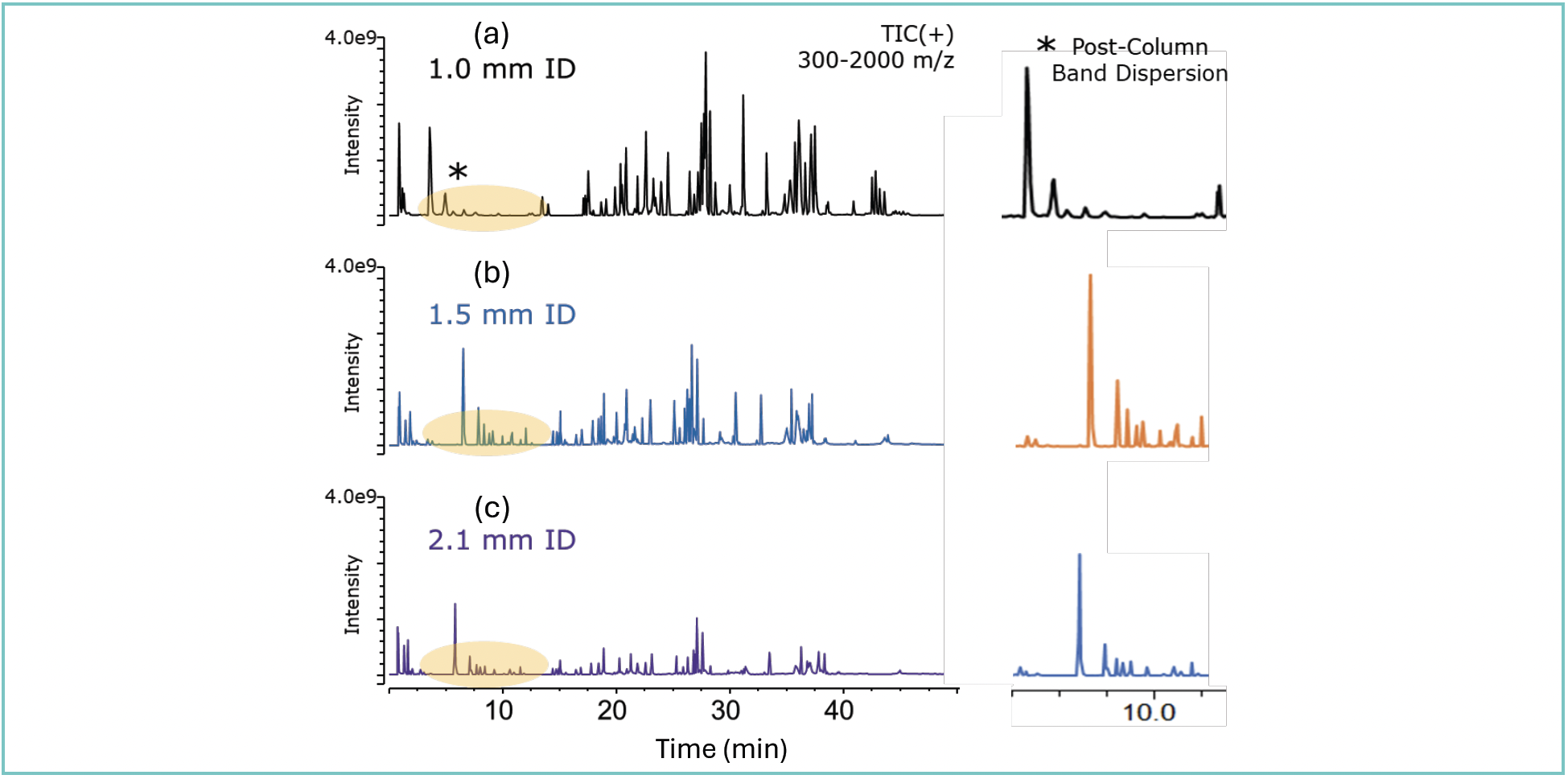
Figure 1 illustrates the effect extracolumn dispersion can play on the separation of peptide fragments from a digested protein. As can be seen from the figure, the 1.5-mm i.d. column provides enhanced sensitivity of the early eluting peptide fragments over both the 1.0- and 2.1-mm i.d. columns. As peak volumes of the late eluting peptides become large enough (relative to instrument dispersion), it does appear that the lower on-column dilution with the 1.0-mm i.d. column has greater impact than the lower impact of instrument dispersion on the larger (1.5-mm i.d.) column. This observation is the topic of further investigation.
FIGURE 1: Effect of extracolumn dispersion on the analysis of digested trastuzumab. Conditions: Column: BIOshell A160 Peptide C18, 15 cm × (a) 1.0-, (b) 1.5-, or (c) 2.1-mm i.d., 2.7 μm; Mobile Phase: [A] water (0.1% [v/v] DFA); [B] acetonitrile (0.1% [v/v] DFA); Gradient: 2 – 50% B in 60 min; Flow Rate: 0.1 mL/min (1.0-mm i.d.), 0.2 mL/min (1.5-mm i.d.), or 0.4 mL/min (2.1-mm i.d.); Column Temp.: 60 °C; Detector: MSD, ESI-(+); Injection: 2.0 μL/min; Sample: trastuzumab tryptic digest, 1.25 mg/mL, 1.5 M guanidine hydrochloride, 0.5% (v/v) formic acid.

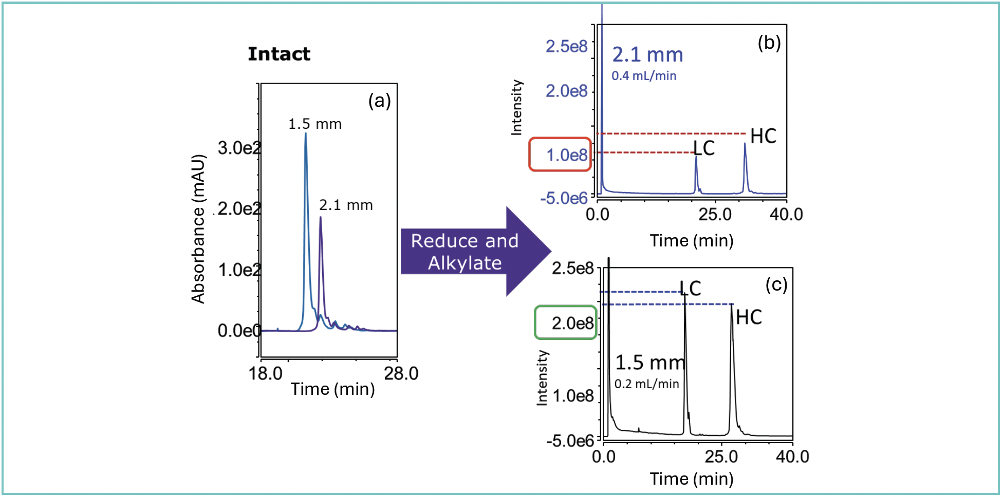
Scaling to lower flow rates is advantageous, especially for large molecule separations. As larger biomolecules have lower diffusion kinetics, lower flow rates are required to achieve maximal diffusion and partitioning between the bulk mobile phase and the stationary phase. Figure 2 illustrates this principle with the analysis of both intact and reduced trastuzumab. In both examples, the 1.5-mm i.d. column led to a doubling of the area counts for both the intact and reduced analysis of trastuzumab. In particular, in the reduced and alkylated analysis, the area counts and sensitivity were improved significantly (2.7x increase in area count for the light chain [LC] and 2.3x increase in area count for the heavy chain [HC]) when using the 1.5-mm i.d. column as compared to a 2.1-mm i.d. column.
FIGURE 2: (a) Analysis of intact and reduced trastuzumab. Conditions: Column: BIOshell IgG 1000 Å Diphenyl, 15 cm × (b) 2.1- or (c) 1.5-mm i.d., 2.7 μm; Mobile Phase: [A] water (0.1% [v/v] DFA); [B] 50:50 acetonitrile (0.1% [v/v] DFA): n-propanol (0.1% [v/v] DFA); Gradient: 27–36% B in 40 min; Flow Rate: as indicated; Column Temp.: 60 °C; Detector: MSD, ESI-(+); Injection: 3.0 μL; Sample: trastuzumab, 1.0 mg/mL, 100 mM ammonium bicarbonate

Oligonucleotides have become an emerging and promising therapeutic modality to treat multiple different diseases (6,7). Owing to the success of the Covid-19 messenger RNA (mRNA) vaccines, these biomolecules have been the subject of renewed research in both medicinal chemistry and biochemistry. As more of these molecules enter the clinical pipeline, the need for robust and sensitive analytical assays grows more urgent. During the process of oligonucleotide synthesis, several impurities can present themselves in the reaction mix including truncated species (n-1, n-2, etc.), species with additional oligonucleotides added to the main sequence (n+1, n+2, etc.), modified oligonucleotides (methylated, oxidized, etc.), or, if phosphorothioate bonds are present, diastereomers of a particular oligonucleotide species. As impurities present in the final drug product may lead to either reduced efficacy or even allergic response in the patient, methods with high sensitivity are necessary.
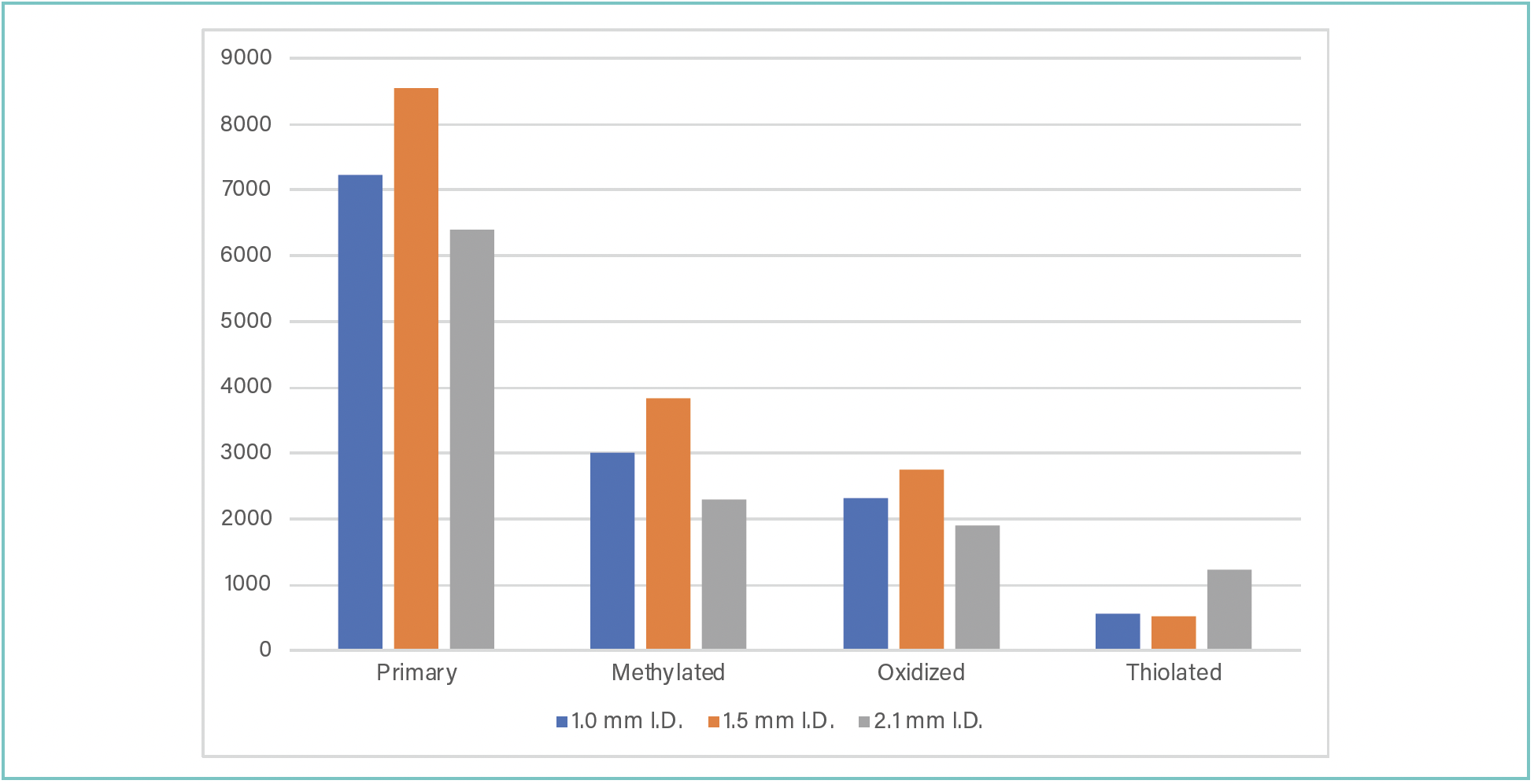
Figure 3 illustrates the differences in sensitivity for the analysis of a “signature” oligonucleotide sequence and its modified variants. As is seen from the bar chart, the 1.5-mm i.d. column achieves sensitivity exceeding that of the 2.1- and 1.0-mm i.d. column on both the primary sequence, methylated sequence, and oxidized sequence. For the thiolated sequence, the 2.1-mm i.d. achieved better sensitivity than the 1.5- or 1.0-mm i.d. columns. This observation may, in part, be due to extensive band broadening seen when analyzing this mixture of diastereomers of the oligonucleotide on the various columns with the narrower internal diameter columns being partially overloaded.
FIGURE 3: Sensitivity (peak height as y-axis) vs. column i.d. (as x-axis): Comparison of peak heights of modified and unmodified oligonucleotides analyzed with the BIOshell A160 Peptide C18 column (15 cm length) with different i.d.’s. Conditions: Column: BIOshell A160 Peptide C18, 15 cm × 2.1-, 1.5-, or 1.0-mm i.d., 2.7 μm; Mobile Phase: [A] water (15 mM triethylamine [TEA], 400 mM hexafluoroisoproanol [HFIP]), [B] water (15 mM TEA, 400 mM HFIP): methanol (40:60 v/v); Gradient: 15 to 50% B in 15 min; Flow rate: 0.1 mL/min (1.0-mm i.d.), 0.2 mL/min (1.5-mm i.d.), or 0.4 mL/min (2.1-mm i.d.); Column temp.: 70 °C; Detector: MSD, ESI-(+); Injection: 1.0 μL; Sample: Oligonucleotide mix; 100 μM, water (10 mM Tris-EDTA, pH 8.0).

Conclusion
Narrow internal diameter columns are advantageous for the analysis of biomolecules as the lower flow rates necessary to achieve optimal chromatographic performance are a requirement in efficiently using columns with very narrow internal diameters. However, larger volumes in the flow path or instrument, poorly packed columns, or incorrect method scaling can lead to a reduction in sensitivity and efficiency. The use of a 1.5-mm i.d. UHPLC column was demonstrated in this paper for the analysis of different classes and sizes of biomolecules. Trastuzumab was analyzed at the intact, middle-up, and bottom-up levels, and in all cases, the 1.5-mm i.d. column enabled higher sensitivity than either 2.1- or 1.0-mm i.d. columns. For the analysis of oligonucleotides and modified oligonucleotides, the 1.5-mm i.d. column again demonstrated improved sensitivity over 2.1- and 1.0-mm i.d. columns with the only exception being the thiolated species. The incorporation of a 1.5-mm i.d. column into the chromatographer’s toolbox is a relatively simple and effective way to get more sensitivity out of analytical assays without the expense of more sophisticated instrumentation.
References
(1) Gritti, F. A Stochastic View on Column Efficiency. J. Chromatogr. A 2018, 1540, 55–67. DOI: 10.1016/j.chroma.2018.02.005
(2) Gritti, F.; Wahab, M. F. Understanding the Science Behind Packing High-Efficiency Columns and Capillaries: Facts, Fundamentals, Challenges, and Future Directions. LCGC N. Am. 2018, 36, 82–98.
(3) Lestremau, F.; Wu, D.; Szuecs, R. Evaluation of 1.0 mm I.D. Column Performances on Ultra High Pressure Liquid Chromatography Instrumentation. J. Chromatogr. A 2010, 1217, 4925–4933. DOI: 10.1016/j.chroma.2010.05.044
(4) Wu, N.; Bradley, A. C.; Welch, C. J.; Zhang, L. Effect of Extra-Column Volume on Practical Chromatographic Parameters of Sub-2 μm Particle-Packed Columns in Ultra-High Pressure Liquid Chromatography. J. Sep. Sci. 2012, 35, 2018–2025. DOI: 10.1002/jssc.201200074
(5) Libert, B. P.; Godinho, J. M.; Foster, S. W.: Grinias, J. P.; Boyes, B. E. Implementing 1.5 mm Internal Diameter Columns Into Analytical Workflows. J. Chromatogr. A 2022, 1676, 463207–463213. DOI: 10.1016/j.chroma.2022.463207
(6) Yin, W.; Rogge, M. Targeting RNA: A Transformative Therapeutic Strategy. Clin. Transl. Sci. 2019, 12, 98–112. DOI: 10.1111/cts.12624
(7) Andersson, S.; Antonsson, M.; Elebring, M.; Jansson-Loefmark, R.; Weidolf, L. Drug Metabolism and Pharmacokinetic Strategies for Oligonucleotide and mRNA-based Drug Development. Drug Discov. Today 2018, 23, 1733–1745. DOI: 10.1016/j.drudis.2018.05.030
ABOUT THE AUTHOR
Cory E. Muraco is the Biomolecule Workflows Manager at MilliporeSigma, the life science business of Merck KGaA. After finishing his graduate studies at Youngstown State University (Youngstown, Ohio, USA), Cory started his career at MilliporeSigma in 2013. After holding several roles in both R&D and Marketing functions, in 2022, Cory assumed his current role as the Biomolecule Workflows Manager where he is tasked with designing, developing, and executing the marketing and R&D strategies around MilliporeSigma’s bioanalytical initiative. Cory is the author of several manuscripts appearing in trade magazines and has delivered over 100 presentations at international conferences, roundtable symposia, and at various pharmaceutical and biopharmaceutical companies. Direct correspondence to: cory.muraco@milliporesigma.com

New Study Investigates Optimizing Extra-Column Band Broadening in Micro-flow Capillary LC
March 12th 2025Shimadzu Corporation and Vrije Universiteit Brussel researchers recently investigated how extra-column band broadening (ECBB) can be optimized in micro-flow capillary liquid chromatography.


