Green Chromatography (Part 3): Sample Preparation Techniques
LCGC Europe
Discussing the latest developments in sample preparation techniques that reduce environmental impact.
The first two articles of this series on green chromatography from the Research Institute for Chromatography (RIC) covered green liquid, gas and supercritical fluid chromatography in practice. Part 3 focuses on sample preparation, potentially the most polluting step of any analytical method because of the high quantities of organic solvents used.
Great efforts have been made to develop sample preparation techniques that guarantee high recovery and reproducibility and that are also greener, faster, cheaper and easier to automate than older methods. Other green trends are on-site or in-field analysis and the development of sample preparation methods that allow multi-residue determination and/or methods in which several sample preparation processes are combined in one step. Portable and transportable GC and GC–MS systems are now commercially available for on-site analysis. All those methods are contributing to green chemistry because the expensive, energy-consuming transport of samples to laboratories is reduced. On-site sampling also has the advantage that remediation or re-sampling can take place immediately after the first analytical results are obtained.

The advent of advanced mass spectrometric (MS) technologies in recent years had several consequences on our analytical work including the sample preparation step. On the one hand, new and more stringent requirements were formulated by regulatory agencies and more and more samples have to be prepared and analysed. On the other hand, because of the high sensitivity and selectivity of mass spectrometers in GC–MS–MS and LC–MS–MS combinations, sample preparation can be simplified and miniaturized. In the past, enrichment was of vital importance in the sample preparation step because samples were too dilute for direct analysis and needed to undergo a chain of specific treatments to make them compatible with the sensitivity of the analytical techniques used. With present state-of-the-art instrumentation, detectable and quantifiable quantities are in the pico- and femtogramme range and the dictum "the best sample preparation is no sample preparation"1 can be re-considered and re-evaluated.
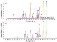
Figure 1: Analysis of pesticides in a spiked surface water by (a) direct LCâMSâMS (b) in-line SPEâLCâMSâMS. Peak numbers (ionization mode, precursor and product ions): (1) Metamitron (+, 203.1, 175.1), (2) Dicamba (â, 221.0, 177.0), (3) Fluroxypyr (â, 253.0, 194.9), (4) 2,4-dinitrophenol (â, 183.0, 108.9), (5) Lenacil (â, 233.1, 150.9), (6) Bentazon (â, 239.0, 132.0), (7) 2,4-D (â, 218.9, 160.9), (8) Bromoxynil (â, 275.9, 78.9), (9) MCPA (â, 199.0, 141.0), (10) DNOC (â, 197.0, 180.1), (11) 2,4-DP (â, 218.9, 160.9), (12) 2,4,5-T (â, 252.9, 194.9), (13) MCPP (â, 213.0, 141.0), (14) Dimethomorph (+, 388.1, 301.0), (15) MCPB (â, 227.0,141.0), (16) Dimethenamide (+, 276.1, 244.0), (17) Tebuconazole (+, 308.2,70.0).
A typical application is the multi-residue determination of pesticides in drinking and surface waters. Figure 1(a) shows the direct analysis of a surface water spiked with 17 target pesticides at 50 ng/L. Before injection, the water sample was acidified with 0.2% formic acid and filtered through a 0.45 µm cellulose filter. The injection volume was 400 µL. The column was a Zorbax Eclipse Plus C18 with 10 cm × 2.1 mm i.d., 3.5 µm particles (Agilent Technologies, Wilmington, Delaware, USA) and a gradient from 10% B to 79% B in 8 min (A = 0.05% formic acid in water, B = acetonitrile) was applied. All spiked pesticides could easily be elucidated and quantified in one run by using alternating positive/negative electrospray ionization with JetStream technology (Agilent Technologies, Santa Clara, California, USA) and multiple reaction monitoring (MRM). Limits of quantification (LOQs) are in the ng/L level for the acidic pesticides (negative mode) and in the subng/L level for the basic pesticides (positive mode). In the 1990s, these sensitivities could only be obtained using labour- and time-consuming sample preparation methods, such as solid-phase extraction (SPE) of a 1 L sample and concentration to 1 mL (enrichment factor 1000), followed by LC-diode array detection (DAD) analysis. Even lower concentrations (ca. tenfold) can be measured by combining SPE placed in-line with LC–MS–MS and using the LC configuration shown in Figure 2. Figure 1(b) shows the analysis of 5 mL of the same water sample spiked at 1/10 compared with Figure 1(a) on the same LC–MS–MS system but equipped with a column switching valve containing up to six Oasis HLB cartridges with 20 mm × 2.1 mm i.d., 25 µm particles (Waters Corporation, Milford, Massachusetts, USA) and an extra pump for loading the cartridges. After sample loading, the cartridge is desorbed using the analytical LC pump and the enriched solutes are analysed. By optimization of LC parameters, peak distortion can be avoided, resulting in very similar resolution to direct water injection, while the sensitivity is increased by a factor of 10 to 12.
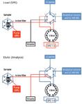
Figure 2: Configuration for on-line SPEâLCâMSâMS; Load (SPE): Sample enrichment onto SPE cartridge, Elute (Analysis): Liquid desorption of the SPE cartridge and LCâMSâMS analysis.
SPE, in its different forms, is nowadays a key component in sample preparation and will be discussed further. It not only removes matrix components that can interfere in MS ion formation (ion suppression) but also selectively enriches the solutes of interest (targets) with a minimum quantity of organic solvent. GC–MS–MS and LC–MS–MS have revolutionized our sample preparation strategies. Systems are now available with complete automated data handling to determine around 600 endocrine disrupting chemicals (EDCs) by GC–MS–MS or more than 300 nonGC amenable pesticides by LC–MS–MS using two transitions and dynamic MRM. We recently described several green sample preparation methods [direct injection, automated off-line SPE and miniaturized liquidliquid extraction (LLE)] that were used in combination with LC–MS–MS for the determination of biogenic amines, mycotoxins, stilbenes, flavonoids, tannins, anthocyanins and phenolic acids in wines and grape juices.2
In the framework of this relatively short contribution on green sample preparation methods, it is impossible to review all achievements made in recent years. We refer the readers to some publications dealing with green sample preparation techniques for environmental analysis3,4 and for food analysis.5 Some new methods will be discussed in depth and illustrated with examples from the laboratories of the authors. A subdivision is made based on the nature of the compounds and the analytical techniques commonly applied for their analysis such as (1) volatile organic compounds (VOCs) that can be analysed by GC via headspace (HS) techniques and (2) semi-volatile organic compounds (SVOCs) that are GC-amenable and nonvolatile (NVOCs) or thermally labile compounds for which LC is the technique of choice.
Last but not least, a number of fully automated sample preparation techniques are described. Automation is very often accompanied with miniaturization and thus solvent reduction.
Volatile Organic Compounds
Gas phase sampling is the greenest technique available since inert gases are used as the extracting phase. Moreover, only volatiles are introduced in the GC instrument and column, guaranteeing minimal maintenance costs and long column lives. Different approaches are available for gas phase sampling and the selection is most often dictated by the sensitivity required. Best known are static headspace (SHS), in-tube extraction (ITEX), dynamic headspace (DHS), purge and trap (P&T), solidphase microextraction (SPME) and headspace sorptive extraction (HSSE). Gas phase sampling is intensively used in environmental and food analysis, and recently in metabolomics (breath analysis).5
In recent years, the applicability of gas phase extraction has been expanded to the analysis of semi-volatiles by performing the extractions at higher temperatures followed by trapping on a sorbent or a cryotrap to focus the solutes before injection. This solventless technique often called gas phase stripping can be applied, for example, to extract polycyclic aromatic hydrocarbons (PAHs) from dust or soil, residual monomers, antioxidants and stabilizers from plastics, etc. An application developed at RIC concerns the determination of aldehydes, hydrocarbons, free fatty acids, vitamins and sterols in vegetable oils. Fractionation and enrichment of these classes out of the apolar triglyceride matrix is a challenging task.
The sampling procedure is as follows: 10 mg vegetable oil placed in a glass microvial is introduced in a thermal desorption unit (TDU). Under a carrier gas flow of 100 mL/min helium, the tube is heated from 25 °C to 250 °C at 60 °C/min and maintained at 250 °C for 20 min. The gas phase extract is focused in a programmed temperature vaporization (PTV) injector at -50°C. The cryotrap is then heated at 12 °C/s to 310 °C (5 min) with a split ratio of 1/10 and a helium flow of 1 mL/min. The products are swept in the column and analysed by GC–MS on a HP-5MS 30 m × 0.25 mm i.d., 0.25 µm df column (Agilent Technologies, Folsom, California, USA) with a temperature programme from 50 °C to 300 °C at 8 °C/min. Figure 3 shows a typical chromatogram for an extra-virgin olive oil batch. Repeatability of thermal extraction analyses is below 10% relative standard deviation (RSD).

Figure 3: GCâMS chromatogram of gas phase stripping of olive oil. Peak numbers: (1) Aldehydes, (2) Fatty acids (palmitic and oleic acid), (3) Squalene, (4) Vitamine E, (5) Sterols.
Based on gas phase extraction, we recently described a novel method for the determination of PAHs in ambient air.7 The method is based on active sampling on sorption tubes consisting of polydimethylsiloxane (PDMS) foam, PDMS particles and a TENAX TA bed. After sampling, the solutes are quantitatively recovered by thermal desorption and analysed by capillary GC–MS. The new sampling method has been compared with the classical method using high-volume sampling on a glass fibre filter followed by polyurethane foam (PUF) for 24 h sampling of ambient air. Volumes enriched were 144 L on the mixed bed and 1296 m3 with the classical method. The latter method required high volumes of dichloromethane to recover the PAHs from the glass fibre and PUF filters while the sorptive method is solventless. The concentrations measured using the new method were significantly higher that the values obtained using the classical method, (i.e., a factor 1.2 to 3 higher for the high-molecular-weight PAHs and up to 35 times higher for naphthalene and 23 times for acenaphthene). The total toxicity equivalence (TEQ) value for PAHs was around two times higher compared with the conventional method, illustrating that the concentrations of PAHs in ambient air have been underestimated until now and that miniaturization of the sample preparation step may result in improved accuracy and precision. In the same framework, we recently illustrated that nitroPAHs, which are present in a concentration range of ca. 100 times lower compared with the PAHs in air, can be determined by GC–MS–MS analysis without any further fractionationconcentration step.8 This is due to the unique specificity characteristics of MS–MS.
Semi-volatile and Non-volatile Organic Compounds
The matrixes in which SVOCs and NVOCs have to be determined include solid and liquid samples. The first step is a solvent extraction step to enrich the target solutes from the matrix. Extraction is commonly followed by a clean-up or fractionation step to selectively remove interferences that disturb the chromatographic or mass spectrometric analysis.
Well known methods for extraction of solutes from solid matrixes include shake flask extraction, Soxhlet extraction (introduced in 1850!), and its modern forms Soxtec and Soxtherm. The techniques often use hazardous solvents for exhaustive extraction. More environmentally friendly techniques include ultrasonic extraction (UE), supercritical fluid extraction (SFE), pressurized liquid and accelerated solvent extraction (PLE and ASE), microwave assisted solvent extraction (MASE) and matrix solid-phase dispersion (MSPD).
UE uses mechanical energy in the form of shearing action that is produced by a low-frequency sound wave. The sample is immersed in an ultrasonic bath with a solvent and subjected to ultrasonic radiation for around 15 min. Advantages over other techniques are simplicity, productivity and low solvent consumption. The excellent properties offered by a supercritical fluid, namely tuneable solvating power (selectivity), high diffusivity and low viscosity, have resulted in analytical usage for many years. The green carbon dioxide (CO2) eventually containing some percentages of methanol or acetone, are most often applied. In recent years enthusiasm on SFE has declined as the disadvantages became clearer. The most important disadvantage is the complexity of the extraction procedure which is strongly matrix dependent and needs careful optimization. A procedure developed for one particular matrix does not automatically work for another matrix. PLE encloses a number of extraction techniques that use solvents at elevated temperatures and pressures. Best known is ASE that originates from SFE and uses organic solvents at high temperature and high pressure to leach out the organics from solid matrixes. Compared with extractions at or near room temperature and at atmospheric pressure, ASE delivers enhanced performance by the increased solubility, the improved mass transfer and the disruption of surface adsorption by the conditions applied. Solvent consumption is typically 1/20 of a conventional Soxhlet extraction. Disadvantages of ASE are the lack of selectivity which means that often further clean-up is needed and that the sample is too dilute for direct analysis and concentration is required.
Recent developments to circumvent these shortcomings are the combination of ASE with SPE and the application of large volume injection (LVI). Another variant of PLE is extraction at high temperatures and pressures with water. Super-heated water extraction (SHWE) including many applications was recently reviewed by Smith.9 Although SHWE is by far the most "green" extraction technique, the main disadvantages of SHWE over ASE are that the solutes are obtained in a dilute aqueous medium and further extraction with an organic solvent is required; that a large number of matrix compounds are extracted as well so that further clean-up is needed; and, last but not least, that the thermal stability of the target solutes under SHWE conditions should be carefully evaluated. MASE uses electromagnetic radiation to desorb organics from their solid matrices. MASE typically operates at 2.45 GHz. In recent years, different systems have become commercially available and they are based on extraction in a closed high pressure vessel with microwave-absorbing solvents, extraction with a non-microwave-absorbing solvent in an open vessel and/or extraction with a non-microwaveabsorbing solvent in a closed vessel applying a micro-wave-energy-absorbing Weflon stir bar (Milestone S.r.l., Sorisole, USA) that heats the solvent. The performance of MASE has been compared with other techniques such as ASE and SFE and similar recoveries were obtained for soil and sediment samples.10 The same disadvantages mentioned for ASE apply to MASE, namely lack of selectivity and the dilution effect. Moreover, care should be taken with solutes that are thermolabile or can rearrange under the influence of electromagnetic radiation.
MSPD first reported by Barker et al.11 is a powerful technique for disrupting and extraction of solid, semisolid and viscous samples. A small amount of sample is homogenized with bulk-bonded silica sorbent (typical ratio 1 to 4) with a glass pestle in a glass mortar. The shearing forces disrupt the structures of the matrix and disperse the sample over the surface of the sorbent. The homogeneous blend of sorbent and sample is then packed in a syringetype cartridge (like a SPE cartridge) followed by elution of targets and interfering compounds (or viceversa) by consecutive washings with appropriate solvents. A variation of MSPD is dispersive SPE (DSPE) as used in the Quick, Easy, Cheap, Effective, Rugged and Safe (named QuEChERS) multi-residue method for the determination of pesticides in different matrices (see further).
In the European Community (EC), 1999 will be remembered as the year of the Belgian dioxin crisis. The dioxins (mainly polychlorinated dibenzofurans or PCDFs) originated from animal feed that was contaminated by used transformer oil and many thousands of food samples had to be analysed on polychlorobiphenyls (PCBs). During the crisis, all possible sample preparation techniques were evaluated and a high throughput "green" method based on UE and MSPD was developed. The implementation of the method was accompanied with validation/reproducibility studies and the analysis of certified samples. Samples were homogenised using a blender. From fat samples (chicken or pork fat) 1 g sample was weighed in a 20 mL vial. From eggs, 3 g egg yolk was taken. For animal feed samples or other meat products, a sample size corresponding to 200–500 mg fat was analysed. To the sample, 2 g anhydrous sodium sulphate and 10 mL petroleum ether were added. Tetrachloronaphthalene or octachloronaphthalene may be added as internal standard (IS) to the sample at this stage although external standardization can also be applied. The vial is closed and placed in an ultrasonic bath at 30 °C for 30 min. In this step, the fat and PCBs are transferred from the matrix in the petroleum ether phase, while sodium sulphate adsorbs the water present in the sample.
After extraction, the sample is allowed to settle. An aliquot (typically 5 mL) is transferred to a test tube and another aliquot (2 mL) is used to gravimetrically determine the fat content of the extract. To the test tube, 2 g of acidic silica gel (44% sulphuric acid) is added and the tube is vortexed for 2 × 30 s. This technique is nowadays called DSPE. In column chromatography and in SPE, the analytes are eluted through a bed and the fat is retained. In DSPE, the fat matrix is allowed to bind on the adsorbent that is mixed with the sample, while the solutes of interest stay in solution. For the fractionation of fat from PCBs, this method works very efficiently. After settlement of the adsorbent or centrifugation, an aliquot of the clear solution is transferred to an autosampler vial. The final volume is not important, because the concentrations are calculated to the initial 10 mL solvent and the sample or fat weight. One technician can easily prepare more than 50 samples a day and the solvent quantity needed is substantially lower (ca. a factor of 20) compared with conventional methods in which, besides extraction, column chromatography has to be performed. The reproducibility and accuracy of the UE–DSPE method was evaluated with the determination of the PCB content in a certified reference material of the EC namely the mackerel oil sample CRM 350 (IRMM, Geel, Belgium). Analyses were performed by four laboratories to which the sample preparation method was transferred and using retention time locked (RTL) GC–MS. The results are summarised in Table 1.

Table 1: Accuracy and reproducibility test for the mackerel oil sample.
Extraction of liquid samples (water samples, beverages, biological fluids, organic solvent extracts obtained by extraction of solid samples, etc.) is based on partitioning into an immiscible extracting phase which can be liquidlike in LLE, gum-like in SPME using a PDMS fibre or solid-like in SPE.
In environmental analysis, LLE is still carried out manually by shaking the water sample with an organic solvent such as dichloromethane or n-hexane in a separation funnel. The volume of the extract is usually too large for direct injection and in order to obtain sufficient sensitivity, additional evaporation-concentration steps are necessary. Miniaturization of LLE has been the subject of intensive research in the last thirty years. In 1981, W. Jennings presented a rapid liquid-liquid extractor for very small solvent/sample volume ratios (typically 20 µL/40 mL) and applied the technique for analysis of an apple distillate concentrate.12 In fact this can be considered as the first application of single-drop microextraction (SDME) (see further). Micro LLE in the vial of an autosampler has been developed in the beginning of the 90s and illustrated for the extraction of haloforms from drinking water.13 Shortly after the introduction by Pawliszyn et al.14 of SPME (in fact liquidphase microextraction when PDMS is used as fibre coating) numerous micro-LLE methods were introduced. Jeannot and Cantwell,15 and He and Lee16 described SDME in 1997. The main disadvantage of the method is drop instability and a development trying to overcome this was liquid phase microextraction (LPME) using a membrane between sample and microdrop.17 A similar approach is used in membrane assisted solvent extraction (MESA) developed by Popp et al.18 and commercially available from Gerstel (Mülheim an der Ruhr, Germany) in a fully automated format. Extraction volumes used are typically 100–150 µL.
In SDME and LPME it can take a relatively long time for equilibration to take place between the aqueous solution and the droplet. This drawback has been addressed by developing a method based on the dispersion of tiny droplets of the extraction liquid within the aqueous solution. The method named dispersive liquid-liquid microextraction (DLLME)19 is in essence close to cloud point extraction (CPE) and can be regarded as multiple-drop microextraction (MDME). In CPE, an involatile surfactant is added while in DLLME a disperser solvent such as diethylether or acetone is used and the extraction medium is a high-density solvent such as tetrachloroethylene. The extraction process can be optimized by using ultrasounds (USA-DLLME or ultrasound- assisted DLLME).20 Notwithstanding the merits of the above techniques, it is very difficult to validate and, moreover, to automate them.
In SPME using PDMS or polyacrylate (PA), partitioning occurs between the gum-like liquid coated on a fibre and the aqueous solution. The analytes are then desorbed from the fibre in-line with the chromatographic system. SPME is a cheap, fully automated solventless technique that is widely used for preparation of food, environmental and bio samples before capillary GC analysis.14 Stir bar sorptive extraction (SBSE) is based on the same principles as SPME but the quantity of extracting solvent (PDMS) is much higher.21 In a typical experiment, a stir bar coated with 25 mg of PDMS is added to 10 to 250 mL of an aqueous solution containing the compounds of interest and extraction is carried out at 1000 rpm during 30 min. The stir bar is then removed from the sample and the analytes dissolved in the PDMS are released by thermal desorption for GC or liquid desoption for LC analysis. Limits down to the ng/L level can easily be reached. Pre-concentration is based on the log P value and for compounds with low log P values in situ derivatization can be applied. As an example, derivatization was used for the determination of bisphenol-A (BPA) in wash water of polycarbonate (PC) baby bottles. In situ derivatization was carried out with acetic acid anhydride. The detection limit was 0.12 ng/L of BPA. PC baby bottles were subjected to simulated every day use and the influence of heating in a water bath and by microwaves has been studied.22
SPE is increasingly being applied for isolating or enriching compounds from aqueous samples and is much faster, cheaper and more versatile than most classical techniques. When properly designed, SPE methods need minimum quantities of organic solvents to recover the targets. Moreover, SPE procedures are easily automated using robotics, or can be coupled on-line with chromatographic techniques. The principle of retention (enrichment) is analogous to LC and is suitable for low, intermediate and high polarity solutes. Commonly, SPE media are based on normal phase (NP), reversed phase (RP) or ion exchange chromatography (IEC). Other specific SPE supports include restricted access media (RAM), immunosorbents and molecularly imprinted polymers (MIPs). Giving an overview of the different SPE sorbents and their applications is not within the scope of this paper. Hundreds of SPE cartridges are commercially available and the catalogues of the manufacturers describe their features and applications. The principles and practice of SPE have been described in the book of Simpson (Ed.).23 The syringe type cartridge, packed with particles of 30 µm to 60 µm, is the most common SPE format. Solvent flow through is typically done using a commercially available vacuum manifold. In recent years, because of the performance of modern mass spectrometers in terms of sensitivity, SPE is intensively used to remove matrix compounds rather than to enrich targets.
A variant of SPE is DSPE. In DSPE the sorbent is added to a solvent extract and unwanted compounds are retained on the sorbent while the target compounds remain in solution. DSPE was intensively used in 1999 in our laboratory for the Belgian dioxin crisis as described before and similar procedures are nowadays used in the QuEChERS multiresidue method.24,25 Today, the QuEChERS method is by far the best developed multi-residue method for a wide variety of pesticides in several common non-fatty and, since recently, fatty food matrices. DSPE is a key aspect of the method and the type of adsorbents, as well as the pH and polarity of the solvent, can be adjusted for differing matrices and difficult analytes. We recently described the determination of 40 pesticides in baby food using a QuEChERS kit for extraction and DSPE (Agilent Technologies) in combination with UHPLC–MS–MS.26 Detection limits were between 500 ng/kg and 10 ng/kg which is much lower than the maximum residue level (MRL) of 10 µg/kg imposed by the European Union. The green character of QuEChERS has recently been highlighted by the inventors of the technique with about 95% reduction in solvent consumption, 95% in consumable costs and 90% reduction in time27 compared with the traditional multi-residue method used in Europe for nearly 20 years. Automated DSPE clean-up for QuEChERS extracts has been developed and has recently become commercially available.28 One of the disadvantages of exhaustive extraction aimed at in QuEChERS is that the extract may still contain involatile matrix material that can have a detrimental effect on the chromatographic performance. This is especially the case when GC is the analytical method. Contaminated inlet liners lower the response of some pesticides affecting the accuracy and precision of the analysis. The performance can be restored by exchanging the inlet liner and/or cutting the inlet part of the column. An automated solution is provided by using an automated liner exchange system (ALEX).29
SBSE is also applied as multi-residue sample preparation method for pesticide analysis.30,31 Compared with QuEChERS, SBSE has the advantage that the DSPE steps are omitted and that higher sensitivity is obtained (all extracted solutes can be quantitatively transferred to the column using thermal desorption). The limitation is that the log P value of the solutes should be at least 2 for good enrichment. A typical example is shown in Figure 4 for the determination of pesticides in a grape sample.
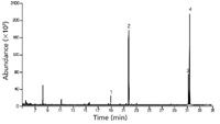
Figure 4: SBSEâGCâMS ion extracted chromatogram (IEC) of pesticides in grapes. Peak numbers: (1) Procymidone, (2) Permethrin I, (3) Permethrin II.
The initial step can be similar to QuEChERS (i.e., extraction of 10 g homogenized sample with 10 mL acetonitrile). For SBSE, 1 mL of the extract is taken and diluted to 10 mL with water. A stir bar 10 mm long coated with 0.5 mm PDMS is added and the mixture is stirred for 60 min at 1000 rpm. After sampling, the stir bar is taken out with tweezers, dipped in distilled water, placed on lintfree tissue to remove residual droplets and finally put in a liner of a thermal desorption system. Splitless thermal desorption was performed by heating the tube from 40 °C to 280 °C (5 min) at a rate of 60 °C/min. The analytes were cryo-focused in a PTV at –150°C with liquid nitrogen prior to injection. An empty baffled liner was used in the PTV injector. For splitless injection (2 min) the PTV was ramped from –150°C to 280 °C (2 min) at a rate of 600 °C/min. Capillary GC analysis was done on a 30 m × 250 µm i.d., 0.25 µm df HP-5MS column (Agilent Technologies). The oven was programmed from 70 °C (2 min) at 25 °C/min to 150 °C, at 3 °C/min to 200 °C and finally at 8 °C/min to 300 °C. Screening of pesticides was performed using the automatic RTL screener software in combination with the Agilent RTL pesticide library. The RTL screener elucidated by ion extraction at m/z = 283 and 183, procymidone (peak 1) and permethrin I and II (peak 2 and 3). The concentration levels were measured at 172 µg/kg (EC norm 5 mg/kg), 20 µg/kg and 83 µg/kg (EC norm for the sum 1 mg/kg), respectively.
Fully Automated Sample Preparation Stations
Downscaling of sample amounts help tremendously not only in solvent savings but also in opening new perspectives for the development of fully automated sample preparation stations off-line or in-line with the chromatographic system.
As an example, we described an automated method for the simultaneous determination of six important organotin compounds in water and sediment samples. The method is based on derivatization with sodium tetraethylborate followed by automated HS-SPME combined with GC–MS under RTL conditions. Deuterated organotin analogues were used as internal standards. The method was validated and excellent figures of merit were obtained. LOQs were from 1.3 to 15 ng/L for water samples and from 1.0 to 6.3 µg/kg for sediment samples.32 The method was successfully transferred to a number of environmental laboratories that received accreditation for organotin analysis in environmental samples. Other laboratories, on the other hand, could not implement the method because local regulatory authorities claimed that the used sample preparation technique, namely SPME, was deviating too much from liquid-liquid extraction described in the official method.33 An automated micro LLE-derivatization method was therefore developed for the determination of the two most important organotin compounds, namely tributyltin (TBT) and triphenyltin (TPhT) in water
All sample preparation steps were automated by using a dual-rail autosampler MPS-2 (Gerstel). Sample and solvent volumes were reduced compared with the official method in order to fulfill the hardware requirements. Figure 5 shows the autosampler equipped with two liquid injection modules (100 µL and 1 mL syringes), two sample trays (2 mL and 20 mL vials), an agitator and two large wash stations for rinsing with ethanol, water and hexane. The GC–MS–MS is equipped with a PTV injector for LVI.
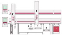
Figure 5: Dual-rail configuration: (1) 20 mL vial tray, (2) 2 mL vial tray, (3) agitator, (4) large wash station (2x), (5) 100 µL LVI syringe, (6) 1 mL syringe.
In the reference method, derivatization is performed in 1 L water that is extracted with 20 mL hexane. The hexane layer is then evaporated to 1 mL. In the automated method, 5 mL water sample is pipetted in a 20 mL vial and 5 mL NaOAc/HOAc buffer (pH 5.3) solution is added. The closed vials are then put in the 20 mL vial tray. The programmed steps are presented in Table 2.

Table 2: Automated sample preparation steps for the analysis of organotin compounds in water
The robot performs the following basic steps: (1) addition of deuterated internal standards and, in case of calibration addition of native organotin compounds, (2) the in-situ derivatization with NaBEt4, (3) LLE with hexane and (4) the transfer of the supernatant to a 1.4 mL sample vial. 50 µL is then injected in the PTV injector. After each addition step, the sample is shaken for a given time and put back into the sample tray. Separation is performed on a HP-5MS capillary column 30 m × 0.25 mm i.d × 0.25 µm df (Agilent Technologies, Folsom) in a temperature programmed run from 50 °C to 300 °C at 10 °C/min. TBT and TPhT together with their deuterated internal standards are measured in MRM-mode using the following precursor and product ions: TBT (291.2 to 178.9 and 291.2 to 122.8), TPhT (351.1 to 196.9 and 351.1 to 119.9), TBT-d27 (318.2 to 189.9 and 318.2 to 125.8) and TPhT-d15 (366.2 to 202.0 and 366.2 to 119.9). Figure 6 shows a typical chromatogram of a water sample spiked with the internal standards at 200 ng/L (ppt) and TeBT-d36 (tetrabutyltin) as surrogate at 400 ng/L. The concentrations of TBT and TPhT were 18 and 21 ng/L, respectively.

Figure 6: Automated analysis of organotin (TBT, TPhT) compounds in a water sample.
A similar dual-rail autosampler was applied for the fully automated analysis of genotoxic sulphonate esters in pharmaceuticals.34 In this application, the drug substance or reaction mixture is diluted in dimethyl sulphoxide-water. A solution of pentafluorothiophenol is added and the methyl-, ethyl- or isopropyl- pentafluorophenylsulphides are formed that can be analysed by static head space gas chromatography mass spectrometry (SHS–GC–MS). To do this, one robot arm is equipped with a liquid syringe for all liquid handling steps and one robot arm is equipped with a headspace syringe for HS sampling and injection in the GC–MS. Excellent repeatability and high sample throughput are obtained, allowing reaction monitoring and product quality control.
Conclusion
Techniques that reduce or eliminate solvent consumption in the sample preparation step have been developed in recent years and are gaining in popularity. Implementation in laboratories is, however, not always straightforward and very often hindered by regulatory directives and timeconsuming (re)validation studies to obtain accreditation.
Nevertheless, implementing these new developments, especially those that can be automated, into routine laboratories is an important step in greening analytical chemistry.
To conclude, let's compare three methods that are currently used for the determination of PAHs in aqueous samples at the 10 ng/L level namely LLE, SPE and SBSE. In LLE, 500 mL water is extracted with 75 mL dichloromethane, the solvent is evaporated, the residue re-dissolved in 1 mL hexane and 2 µL, corresponding to 10 pg on the column, is analysed by GC–MS. In SPE, 100 mL water is sent over a cartridge packed with octadecylsilica or polystyrenedivinylbenzene particles and the PAHs are recovered in 5 mL acetonitrile or iso-octane. The solvent is evaporated and the residue reconstituted in 0.2 mL hexane and 2 µL, corresponding to 10 pg on the column, is analysed. In SBSE, a PDMS stir bar is added to 5 mL water and then thermally desorbed on-line with the GC–MS system. Quantity introduced is 50 pg on the column. The latter technique not only is solvent free, but (i) more sensitive, (ii) stir bars can be re-used for at least 50 samples and (iii) figures of merit are excellent.
Acknowledgement
We would like to thank IWT–Vlaanderen (SBO-project 050178) for financial support of our research activities related to green analytical chemistry.
Pat Sandra is director of the Research Institute for Chromatography (RIC), Kortrijk, Belgium, professor in Separation Science at the Ghent University, Belgium and director of the Pfizer Analytical Research Centre-UGent, Belgium.
Christophe Devos is researcher specialized in sample preparation and automation for GC–MS analysis at RIC, Kortrijk, Belgium.
Bart Tienpont is head of the Instrument Development division at RIC, Kortrijk, Belgium.
Gerd Vanhoenacker is head of the LC and LC–MS division at RIC, Kortrijk, Belgium.
Frank David is R&D Manager at RIC, Kortrijk, Belgium and extraordinary professor at the Ghent University, Belgium.
References
1. P. Sandra, Water Analysis, Publ. Hewlett-Packard, 5962-6216E, 11 (1994).
2. E. Dumont et al., Suppl. AgroFOOD Industry Hi-Tech, 2(3), 13–16 (2010).
3. J. Curylo, W. Wardencki and J. Nasmiesnik, Polish J. Environ. Stud., 16(1), 5–16 (2007).
4. M. Tobiszewski et al., TrAC, 28(8), 943–951 (2009).
5. P. Sandra, F. David and G. Vanhoenacker, in Comprehensive Analytical Chemistry, Food Contaminants and Residue Analysis, (Ed. Y. Pico), Vol. 15, pp.131–174 (2008).
6. C. Robroeks et al., Pediatric Research, 68(1), 75–80 (2010).
7. E. Wauters et al., J. Chromatogr. A., 1190(1–2), 286–293 (2008).
8. F. David and M. Klee, AN 5990-3366EN, Agilent Technologies (2008).
9. R.M. Smith, Anal. Bioanal. Chem., 385(3), 419–421 (2006).
10. J. Dean, Extraction Methods for Environmental Analysis, Wiley, Chichester, 1998.
11. S.A. Barker, A.R. Long and C.R. Short, J. Chromatogr., 475(1–2), 353–361 (1989).
12. W. Jennings, Sample Preparation for Chromatographic Analysis, Hüthig, Heidelberg, 51 (1983).
13. I. Temmerman et al. AN 228-135, Hewlett Packard (1991).
14. J. Pawliszyn, Solid Phase Microextraction. Theory and Principles, Wiley-VCH, New York, 1997.
15. M.A. Jeannot and F.F. Cantwell, Anal. Chem., 69(2), 235–239 (1997).
16. Y. He and H.K. Lee, Anal. Chem., 69(22), 4634–4640 (1997).
17. L. Zhao and H.K. Lee, Anal. Chem, 74(11), 2486–2492 (2002).
18. M. Schellin and P. Popp, J. Chromatogr. A, 1020(2), 153–160 (2003).
19. M. Rezaee et al., J. Chromatogr. A, 1116(1–2), 1–9 (2006).
20. A. Saleh et al., J. Chromatogr. A, 1216(39), 6673–6679 (2009).
21. E. Baltussen, C. Cramers and P.J.F. Sandra, Anal. Bioanal. Chem., 373(1–2), 3–22 (2002).
22. N. De Coensel, F. David and P. Sandra, J. Sep. Sci., 32(21), 3829–3836 (2009).
23. N.J.K. Simpson, Solid Phase Extraction: Principles, Techniques and Applications, Marcel Dekker, New York, 2000.
24. M. Anastassiades et al. J. AOAC Int., 86(2), 412–431 (2003).
26. G. Vanhoenacker, F. David and P. Sandra, AN 5990-5028EN, Agilent Technologies (2009).
27. S.J. Lehotay, M. Anastassiades and R.E. Majors, LCGC Europe, 23(8), 418–429 (2010).
28. V. Settle et al., AN 4/2010, Gerstel, Germany (2010).
29. O. Lerch, AN 7/2010, Gerstel, Germany (2010)
30. P. Sandra, B. Tienpont and F. David, J. Chromatogr. A, 1000(11), 299–309 (2003).
31. N. Ochiai et al., J. Sep. Sci., 28(9–10), 1083–1092 (2005).
32. C. Devos et al., J. Chromatogr. A, 1079(1–2), 408–414 (2005).
33. European Directive EN 17353. Determination of Organotin Compounds.
34. K. Jacq et al., J. Pharm. Biomed. Anal., 48(5), 1339–1344 (2008).
Characterizing Plant Polysaccharides Using Size-Exclusion Chromatography
April 4th 2025With green chemistry becoming more standardized, Leena Pitkänen of Aalto University analyzed how useful size-exclusion chromatography (SEC) and asymmetric flow field-flow fractionation (AF4) could be in characterizing plant polysaccharides.

