Electromembrane Extraction: The Use of Electrical Potential for Isolation of Charged Substances from Biological Matrices
LCGC North America
How the combination of an electroextraction with hollow-fiber liquid-phase microextraction can lead to a selective, rapid new method.
Recently, efforts have been devoted to the miniaturization of existing liquid–liquid extraction methods to make them more versatile and powerful. Reducing the use of hazardous organic solvents due to environmental and cost concerns, time reduction, ease of automation, possible online coupling, high-throughput capability, and the small amounts of matrix available are major incentives that have motivated scientists working towards miniaturization. In bioanalysis, which in this context is defined as the analysis of small drug molecules and their metabolites in biological samples, the complexity of the matrices requires selective and specific sample preparation methods to isolate the analytes of interest. Macromolecules, salts, cellular material, fat, or lipids in biological matrices such as urine, plasma, whole blood, and breast milk can disturb the separation and data analysis steps. In addition, the analytes of interest can often exist in low concentrations (pg/mL–µg/mL). Therefore, a sample preparation method with high degree of selectivity and enrichment is crucial for a successful analysis.

Ronald E. Majors
With these considerations kept in mind, a totally new approach to sample preparation was proposed in 2006 (1). The innovative concept, called electromembrane extraction (EME), combined the technical setup for hollow-fiber liquid-phase microextraction (HF-LPME) (2,3) with known principles for electroextraction (4–10). This combination offers a highly selective sample preparation method using simple equipment and gains a high degree of enrichment within a short period of time.
The EME method extracts charged substances from a small sample volume through a thin membrane of organic solvent immobilized in the wall of a hollow fiber and into a receiver solution inside the lumen of the hollow fiber. This extraction process is forced by an applied potential difference across the membrane, and this combination of well-known liquid–liquid extraction processes with electrokinetic migration yields a rapid and selective sample preparation method for ionic substances. EME has shown to be compatible with a wide range of biological matrices — for example, plasma, whole blood, urine, and breast milk — preparing clean extracts in a short period of time with simple and inexpensive equipment.
This installment of "Sample Prep Perspectives" is a minireview of the EME technique and shows the investigation of several parameters that affect recovery of charged analytes. In addition, a number of application examples will illustrate its potential for the successful extraction of drugs from complex matrices.
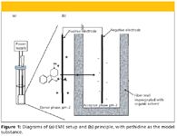
Figure 1
Experimental
The technical setup for the equipment used in EME is based upon earlier experience with HF-LPME and is shown in Figure 1a. The hollow fiber used is made of porous polypropylene, which is compatible with a broad range of organic solvents. The thickness of the wall of the hollow fiber is 200 µm, with a pore size of 0.2 µm and an internal diameter of 1.2 mm. A piece of the hollow fiber is cut to a length of 25 mm and mechanically closed at the lower end with a pair of pincers. The upper end is sealed to the end of a 22-mm pipette tip by heating. To make the supported liquid membrane (SLM), the fiber is dipped in an organic solvent for 5 s to fill the pores in the walls, and the excess of organic solvent gently removed with a medical wipe.
The fiber connected to the pipette tip is guided through a punched hole in the sample compartment cap as illustrated in Figure 1a. The pipette tip works as a mechanical support for a 0.5-mm-thick platinum wire placed inside the lumen of the hollow fiber. Another platinum wire is introduced directly into the donor phase through the sample compartment cap. When coupled to a power supply, these inert wires act as electrodes, thus creating an electrical field across the SLM. In this way, the equipment makes a closed electrical circuit, where the SLM functions as a resistor.
The volume of the sample varies between 150 µL and 500 µL, depending upon the sample compartment size. However, the compartment is never filled more than half full because of the need for convection space. The sample is shaken on a platform shaker during the extraction to increase the physical movement of the analytes in the bulk donor phase and to reduce the thickness of the stagnant layer at the interface between the donor phase and the SLM. The acceptor phase volume is set to 25 µL and is introduced into the lumen of the hollow fiber by a microsyringe. When the predetermined extraction period is finished, 20 µL of the acceptor phase is collected by the microsyringe and transferred to a vial for analysis in a capillary electrophoresis (CE) instrument (11–15) or by high performance liquid chromatography (HPLC) (16).
Results and Discussion
Extraction chemistry: The major driving force in EME is the potential difference across the SLM, in which charged analytes are drawn from the donor phase, across the interface between the donor phase and the organic membrane, through the organic membrane, across the interface between the organic membrane and acceptor phase, and toward the electrode of opposite charge in the acceptor phase. The principle of the extraction is shown in Figure 1b. To ensure full ionization of the model analytes, the pH in both the donor phase and the acceptor phase should be well controlled. In the extraction of basic analytes from water, the pH in the aqueous donor phase was adjusted to 2.0 with 10 mM hydrochloric acid (11–16). The acceptor phase also consisted of 10 mM hydrochloric acid, which increased the solubility of the analytes in the aqueous solution compared to the SLM. In the case of acid extractions, the analytes were dissolved in 10 mM sodium hydroxide to a final pH of 12.0, which also was the pH in the acceptor phase (17).
Choice of organic solvent: The properties of the organic solvent impregnated in the SLM are important for a functional EME system. Based upon earlier experiences with HF-LPME, the nitro compound 2-nitrophenyl octyl ether (NPOE) was tested as the organic solvent for extraction of basic drugs with different polarities. An electropherogram representing the extract after EME of five hydrophobic basic drugs with NPOE as the SLM is shown in Figure 2. Hydrophilic model analytes (log P < 1.0) were unable to penetrate the interface between the donor phase and the SLM with pure NPOE. The high polarity of these analytes with low partition into the SLM seemed to counteract the influence of the electrical field.
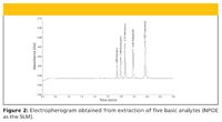
Figure 2
To increase the solubility of the analytes in the organic solvent, di-(2-ethyl-hexyl) phosphate (DEHP) was added to the SLM as an ion-pair reagent (12). The results in Table I illustrate the importance of the nature of the organic solvent. The addition of 10% DEHP to the membrane increased the recoveries of the hydrophilic drugs tested from zero up to 22–54%, while the recoveries of the more hydrophobic analytes tested were reduced dramatically with DEHP presented in the SLM. Also, the hydrophobic drugs ion-paired with DEHP, and were efficiently transported into the organic membrane. However, these ion pairs were highly hydrophobic, and the strong interaction partly prevented them from liberating to the acceptor phase. The addition of 10% tris-(2-ethylhexyl) phosphate (TEHP) to NPOE altered the situation, as shown in Table I. Despite a similar structure to DEHP, TEHP did not function as an ion-pair reagent, as it does not contain any ion-pairing seats. The hydrophilic drugs were not extracted by the addition of TEHP to the SLM. However, the recoveries of the hydrophobic drugs increased with TEHP in the SLM, probably caused by alteration of the polarity in the SLM. A combination SLM consisting of 10% DEHP and 10% TEHP in NPOE led to extraction of almost all the basic drugs tested, showing that development of a membrane that permitted electrokinetic transport of drug substances in a very large logP window was possible.

Table I: The influence of organic solvent on recovery
Proper organic solvents for extraction of acids have been examined (17). The aromatic nitro compounds were found to be inefficient for the acidic model analytes, and aliphatic alcohols with different chain length were tested based upon experiences with HF-LPME (18). The alcohols 1-octanol and 1-heptanol were found to be suitable solvents because of their insolubility in water and easily impregnation in the fiber. In addition, the alcohols showed an appropriate electrical resistance to the applied voltage — that is, the current was low enough to avoid bubble formation and excessive electrolysis but still sufficiently high for the system to maintain the properties of a closed circuit. Recoveries between 8% (hexobarbital) and 100% (fenoprofen) were obtained with 1-heptanol as the SLM (Table II).
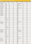
Table II: List of substances and matrices tested in EME
The effect of different organic solvents was strongly dependent upon the potential difference applied across the SLM. The optimal potential difference with NPOE as the SLM was 300 V, which showed recoveries between 16% (pethidine) and 78% (methadone) for the nonpolar model analytes (12). To reduce the applied potential difference, other organic solvents were tested (14). Different SLMs were made with different nitro compounds including ethyl nitrobenzene (ENB) and 1-isopropyl-4-nitrobenzene (IPNB). It was shown that 10 V was the potential difference that showed the highest recoveries, which were in the ranges of 54–80% (ENB) and 59–93% (IPNB). The results show that the chemical nature of the organic solvent in combination with a proper potential difference can be used to design very selective extractions of different analyte classes.
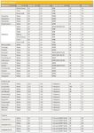
Table II: continued
Other parameters that were found to influence the recoveries in EME included the convection speed, the ion balance between the donor and acceptor phase and the temperature (16).
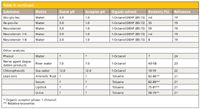
Table II: continued
Extraction by batteries: Because of the successful extractions with only 10 V as the driving power, a simple 9-V battery was tested as the only power supply (14). This replacement of the power supply did not influence the recoveries of the drugs tested, making the idea of a portable sample preparation device possible.
Comparison with HF-LPME: The original idea for EME was to employ the same setup that was used for HF-LPME. The only technical difference between these two systems was the electrical circuit introduced in the EME setup. The platinum electrodes coupled to a power supply created a potential difference across the SLM, hence, changing the extraction mechanism from passive diffusion to electrokinetic migration. A systematic study of the differences between the two methods was performed regarding to the extraction mechanisms, extraction times, recoveries, and selectivity (11). Overall, the introduction of another transport force into the EME system made a significant change in the extraction time compared to the existing HF-LPME model. Generally, the most time-consuming step in bioanalysis is the sample preparation step. Therefore, the extraction speed is of great importance in development of a new extraction method. The time to reach a steady-state level is determined by different parameters in the driving force of the extraction. The only differences between the two systems were the pH in the donor phase, which was 2.0 in the EME system and 12.0 in the HF-LPME system, and the voltage applied across the SLM in the EME system. The recoveries as a function of the extraction times were examined, and the extractions were completed when a steady state level was reached. The graphs in Figure 3 compare the results from an EME extraction with an HF-LPME extraction of the same compound, haloperidol. The time-saving effect obtained from the voltage application in EME was significant, with a reduction in the equilibrium time from about 60 min with passive diffusion as the driving force to 5–10 min with an electrical field as the major driving force. The speed advantage EME offered compared to HF-LPME was most remarkable using the smallest sample compartment tested (300 µL, 31 mm × 4 mm i.d.). In the small compartment, the short electrode distance created a relatively high electrical field strength, resulting in a stronger driving force and hence increased extraction speed.
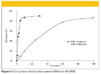
Figure 3
EME from biological matrices:A number of biological matrices have been tested in combination with EME. The matrices covered a wide range of complexity from urine, to diluted and untreated plasma, to whole blood, to breast milk. We will now look at some of these examples.
Human plasma: Extractions of basic drugs from both diluted plasma (14) and undiluted plasma (13) has been tested. These experiments showed that the extraction recoveries were almost independent of the dilution and the pH in the plasma. In other words, extraction from plasma samples without any pretreatment was possible.
The time to reach a steady-state level from untreated plasma was somewhat extended compared to extraction from phosphate buffer (pH 7.4), as exemplified with nortriptyline in Figure 4. The slow extraction kinetics in plasma was probably caused by two factors: the increased viscosity of plasma samples, and drug interactions with plasma proteins. To check whether the viscosity was a deciding factor or not, the plasma sample was diluted with pure water in the ratio 1:1 in a subsequent experiment. The results are shown in Figure 4. It can be seen that lowering the viscosity of the plasma samples did not increase the extraction speed significantly. The steady-state recoveries were comparable in phosphate buffer as well as untreated and diluted plasma.
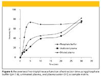
Figure 4
Whole blood: Experiments with EME from whole blood samples have been performed (13). The recoveries obtained from extraction from untreated whole blood spiked with some basic drugs were in the range 19–51%. They were comparable to EME from untreated plasma, even though whole blood is a more viscous and complex matrix than plasma. In some subsequent experiments, whole blood samples were diluted with pure water in the ratio 1:1 or 1:3 to test if a less viscous matrix would increase the recoveries. For the majority of the model analytes tested, the dilution step led to improved recoveries.
Urine: EME of diluted urine samples has been performed (14). The recoveries obtained after 5 min EME from diluted urine were in the range of 37–50%, which were significantly lower than EME from pure water samples under otherwise identical conditions. To test the effect of variety in the water and salt content, urine samples from different persons and collected at different times were spiked with the basic model analytes and subjected to EME. The results showed no significant difference between the recoveries.
Breast milk: EME has been performed on breast milk (14). The recoveries of the drugs tested were in the range 39–55% from breast milk, and the resulting electropherograms showed very few interferences. Extractions from samples collected from two different women and from the start and end of lactation showed no significant differences in the recoveries.
Other applications: The repeatability for the extraction from human plasma was tested at two different concentration levels. The repeatability of the extractions was 5–17% RSD (13). Other EME applications have been performed simultaneously to the works briefly described in this article. EME of peptides (19,20), lead ions (21), chlorophenols (22), nerve agent degradation products (23), and phenols (24) have shown the potential of the technique. Table II gives a comprehensive overview of the substances and matrices that are exposed to EME. The usage of electrical potential difference in sample preparation is also described by Arrigan and his group (4–7). Their research focuses on the electrochemical properties of the interface between two immiscible electrolyte solutions (ITIES), which can be used in separation and detection of ionic molecules from biological matrices.
Conclusion
A new idea about an electrical field as the extraction driving force combined with existing hollow-fiber extraction technology is described briefly. It is shown that the technique offers rapid and selective isolation and enrichment of ionic substances. The extraction recoveries are strongly dependent upon the nature of the organic solvent. This dependence makes EME a highly selective extraction method that uses the nature of the organic solvent and the applied potential to control the extractions.
The time-saving effect by using an electrical field as the driving force is demonstrated during a comparison between EME and liquid-phase microextraction. The potential of EME is shown during extraction from different biological matrices. Extractions from diluted urine, diluted and untreated plasma, diluted and untreated whole blood, and diluted breast milk are noticed, showing promising results. Normally, existing liquid–liquid extraction methods require pH adjustment of the matrix before extraction of ionic analytes. In that way, EME offers possible advantages, where ionic substances can be extracted directly from untreated biological samples without disturbing the chemistry in the sample.
The high extraction speed and the simple equipment needed makes EME a promising tool for field sampling, either from blood samples at the bedside in a hospital or in environmental analysis. The EME experiments performed with a common battery as the only power supply were promising in that respect.
Ronald E. Majors "Sample Prep Perspectives" Editor Ronald E. Majors is business development manager, Consumables and Accessories Business Unit, Agilent Technologies, Wilmington, DE, and is a member of LCGC's editorial advisory board. Direct correspondence about this column to "Sample Prep Perspectives," LCGC, Woodbridge Corporate Plaza, 485 Route 1 South, Building F, First Floor, Iselin, NJ 08830, e-mail lcgcedit@lcgcmag.com.
References
(1) S. Pedersen-Bjergaard and K.E. Rasmussen, J. Chromatogr., A 1109, 183–190 (2006).
(2) S. Pedersen-Bjergaard and K.E. Rasmussen, Anal. Chem. 71, 2650–2656 (1999).
(3) S. Pedersen-Bjergaard and K.E. Rasmussen, J. Chromatogr., A 1184, 132–142 (2008).
(4) D.W.M. Arrigan, Anal. Lett. 41, 3233–3252 (2008).
(5) A. Berduque and D.W.M. Arrigan, Anal. Chem. 78, 2717–2725 (2006).
(6) A. Berduque, J. O'Brien, J. Alderman and D.W.M Arrigan, Electrochem. Commun. 10, 20–24 (2008).
(7) A. Berduque, A. Sherburn, M. Ghita, R.A.W. Dryfe, and D.W.M. Arrigan, Anal. Chem. 77, 7310–7318 (2005).
(8) E. Vandervlis, M. Mazereeuw, U.R. Tjaden, H. Irth, and J. Vandergreef, J. Chromatogr., A741, 13–21 (1996).
(9) E. Vandervlis, M. Mazereeuw, U.R. Tjaden, H. Irth, and J. Vandergreef, J. Chromatogr., A 712, 227–234 (1995).
(10) E. Vandervlis, M. Mazereeuw, U.R. Tjaden, H. Irth, and J. Vandergreef, J. Chromatogr., A 687, 333–341 (1994).
(11) A. Gjelstad, T.M. Andersen, K.E. Rasmussen, S. Pedersen-Bjergaard, J. Chromatogr., A 1157, 38–45 (2007).
(12) A. Gjelstad, K.E. Rasmussen, and S. Pedersen-Bjergaard, J. Chromatogr., A 1124, 29–34 (2006).
(13) A. Gjelstad, K.E. Rasmussen, and S. Pedersen-Bjergaard, Anal. Bioanal. Chem. 393, 921–928 (2009).
(14) I.J.O. Kjelsen, A. Gjelstad, K.E. Rasmussen, and S. Pedersen-Bjergaard, J. Chromatogr., A 1180, 1–9 (2008).
(15) T.M. Middelthon-Bruer, A. Gjelstad, K.E. Rasmussen, S. Pedersen-Bjergaard, J. Sep. Sci. 31, 753–759 (2008).
(16) A. Gjelstad, K.E. Rasmussen, and S. Pedersen-Bjergaard, J. Chromatogr., A 1174, 104–111 (2007).
(17) M. Balchen, A. Gjelstad, K.E. Rasmussen, and S. Pedersen-Bjergaard, J. Chromatogr., A 1152, 220–225 (2007).
(18) T.S. Ho, S. Pedersen-Bjergaard, and K.E. Rasmussen, J. Chromatogr. Sci. 44, 308–316 (2006).
(19) M. Balchen, L. Reubsaet, and S. Pedersen-Bjergaard, J. Chromatogr., A 1194, 143–149 (2008).
(20) M. Balchen, T.G. Halvorsen, L. Reubsaet, and S. Pedersen-Bjergaard, J. Chromatogr., A 1213, 14–17 (2009)
(21) C. Basheer, S.H. Tan, and H.K. Lee, J. Chromatogr., A 1213, 14–18 (2008).
(22) J. Lee, F. Khalilian, H. Bagheri, and H.K. Lee, J. Chromatogr. A 1216, 7687–7693 (2009).
(23) L. Xu, P.C. Hauser, and H.K. Lee, J. Chromatogr., A 1214, 17–22 (2008).
(24) Y.G. Guo, Y.Z. Yu, X.J. Liu, and E.Q. Tang, Chem. Lett. 37, 1272–1273 (2008).
Astrid Gjelstad is with the Department of Pharmaceutical Chemistry, School of Pharmacy, University of Oslo, Oslo, Norway. She defended her PhD thesis about Electromembrane Extraction October 2009, and is continuing her research regarding microextraction and membrane technology in Oslo. astrid.gjelstad@farmasi.uio.no
New Method Explored for the Detection of CECs in Crops Irrigated with Contaminated Water
April 30th 2025This new study presents a validated QuEChERS–LC-MS/MS method for detecting eight persistent, mobile, and toxic substances in escarole, tomatoes, and tomato leaves irrigated with contaminated water.
University of Tasmania Researchers Explore Haloacetic Acid Determiniation in Water with capLC–MS
April 29th 2025Haloacetic acid detection has become important when analyzing drinking and swimming pool water. University of Tasmania researchers have begun applying capillary liquid chromatography as a means of detecting these substances.

.png&w=3840&q=75)

.png&w=3840&q=75)


.png&w=3840&q=75)


.png&w=3840&q=75)


