Dried Blood Spots and High-Resolution Mass Spectrometry for Discovery Fast PK Bioanalysis
Special Issues
This article introduces the advantages of accurate mass high-resolution mass spectrometry LC–MS (HRMS) coupled to the dried blood spot (DBS) technique for fast PK applications in a discovery environment. Compared with the established norm of plasma bioanalysis using triple quadrupoles, HRMS coupled to DBS is a viable alternative. The benefit is access to critical new information (HRMS bioanalysis) and significantly less stress on the animal (DBS), both factors that potentially improve the quality of early PK data.
The use of dried blood spots (DBS) as a sampling technique was pioneered by Dr. Robert Guthrie at the University of Buffalo in the 1960s. The challenge he faced was to obtain blood samples from newborns to screen for phenylketonuria, a genetic metabolism disorder. Dr. Guthrie used a filter paper disk to sample blood via a heel-prick, which he then could place directly atop an agar culture plate used to screen for phenylketonuria (1). Today, the DBS technique is used routinely by hospitals in the developed world to screen newborns for inherited metabolic disorders and its success is related to several practical advantages; the DBS technique is less invasive, requires smaller sample volumes, is easier to transport and store, and reduces risk due to infectious pathogens (2,3). Recently, the pharmaceutical industry has been debating the utility of DBS for pharmaceutical analysis versus the established norm of plasma bioanalysis, primarily in an effort to reduce drug development costs.
In discovery fast pharmacokinetics (PK), however, the advantages are straightforward, especially in terms of animal welfare (4,5). A key advantage is reduced cumulative stress on the animal. There are several factors that can stress the animal and therefore influence the PK profile; bruising, hemorrhaging, and thrombosis resulting from animal handling and venipuncture, surgical implantation of the cannulas for both dosing and sampling, and the associated adverse effects such as blockage, accidental removal, and infection, are all stress factors on the animal. Sampling smaller amounts of blood volume by using the DBS sampling technique has the potential advantage of dramatically reducing the stress factors on the animal, ultimately resulting in better PK data.
One current limitation of the DBS technique is its amenability to automation when compared to established liquid-handling robotics, which make samples ready for analysis by liquid chromatography–mass spectrometry (LC–MS). Robotic liquid handlers enable complete workflow automation of plasma sample clean-up using protein precipitation techniques, allowing a large number of samples to be analyzed simultaneously. In most laboratories, a sample aliquot from a DBS card requires a manual step of coring the center of the dried sample on the card, a step that represents a bottleneck for high-throughput applications. In discovery PK applications in which a multitude of compounds are analyzed every day, tuning of the triple-stage quadrupole MS system for the multiple reaction monitoring (MRM) step specific for the compound of interest represents another step where throughput improvements can be made. While some companies have robotic platforms for DBS work, they are still first generation and do not compete with liquid-handling robotic platforms in terms of efficiency. This article introduces the advantages of accurate mass high-resolution LC–MS (HRMS) coupled to DBS sampling for fast PK applications in a discovery environment to overcome the transition from plasma bioanalysis to a DBS-based workflow.
Experimental
Verapamil (Sigma Chemicals, St. Louis, Missouri) was administered orally and intravenously to catheterized male rats at doses of 2 mg/kg (in 0.5% carboxymethylcellulose) and 1 mg/kg (in 10% hydroxypropyl-β-cyclodextrin), respectively. Blood was heparinized and collected at various timepoints, and immediately spotted in 30-μL aliquots to FTA DMPK-C cards (GE Whatman, Florham Park, New Jersey) via air-displacement pipette. The spots were dried overnight under ambient conditions.
Verapamil standards and QC samples were prepared in fresh heparinized whole blood from spiking solutions. Spiking solutions for standards and QC samples were prepared by serial dilution from independent stock solutions (all in methanol). Pools were immediately spotted as described above and allowed to dry.
Punches (6-mm) were removed from each spot and extracted in 90% methanol–10% water using diphenhydramine as an internal standard. The supernatants were analyzed by LC–MS (HRMS, Exactive, Thermo Fisher Scientific, Madison, Wisconsin) using a resolution setting of R = 25,000, which resulted in a scan speed of 4 scans/s. The mass spectrometer was verified to be in calibration by using a mixture of Ultramark caffeine, and MRFA, but no other optimization was done. A model 1100 high-pressure liquid chromatograph (Agilent Technologies, Palo Alto, California) coupled to a CTC Pal autosampler (Leap Technologies, Carrboro, North Carolina) was used to perform the chromatography and inject the sample.

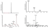
Figure 1: Extracted ion HRMS chromatogram of verapamil (m/z 455.2892) at R = 25,000 (left panel top), and the verapamil spectrum at 1.61 min (right top panel). Total ion chromatogram (bottom left panel) and chemical noise at 1.61 min (bottom right panel).
Results and Discussion
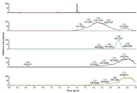
Figure 1 shows the HRMS chromatogram and mass spectra of verapamil from the 6-mm core. While a resolution of R = 25,000 provides more than adequate specificity (right top panel, Figure 1), the presence of coeluted peaks can be seen easily due to the full-scan HRMS acquisition, indicating the extent of clean-up achieved by protein precipitation as a clean-up technique (right bottom panel, Figure 1). The advantages of full-scan HRMS become apparent over the traditional triple-quadrupole–based MRM assay, and one of these advantages is that every sample injected on the HRMS system can be monitored for ion suppression. The data file can be queried postacquisition (Figure 2), and in this example, the data file was queried for the ions (m/z 494, 522, 786, 804) representing the commonly suspected phospholipids. This cannot be done pragmatically on a triple-quadrupole system and requires a separate experiment to characterize the extent of ion suppression. During bioanalysis performed using MRM, it is simply assumed that the components responsible for ion suppression are not coeluted. However, when there is a discrepancy in the bioanalysis, the MRM data file is devoid of any information indicative of what might have caused the variance. Using HRMS, every sample injected can be queried for known components, and acquired data file mined for unknown causes of variance due to the full scan MS nature of the data acquisition.

Figure 2: Postacquisition extracted ion chromatograms of common phospholipids and lysophospholipids displayed relative to the retention time of verapamil, showing the advantage of full-scan HRMS, as every injection can be monitored for interferences.
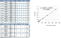
The 6-mm core size provided adequate sensitivity (1 ng/mL) for the parent. The standard curve between the range of 1 ng/mL to 500 ng/mL, with QC samples at 5 ng/mL, 50 ng/mL and 250 ng/mL was linear (r2 of 0.999) and the results are shown in Figure 3. The results indicate that the HRMS system operating in the full-scan mode meets the basic precision and accuracy requirements of non-GLP bioanalysis. Note that the QC levels represent a second independent weighing, used to substantiate the results obtained from the calibration curve (calibrants weighed independently), and have a precision result of less than 6% across the range. This is standard practice put in place to ensure integrity in a semihigh-throughput bioanalytical operation.

Figure 3: Calibration curve results in the range 1â500 ng/mL including QC samples at 5, 50, and 250 ng/mL using the Exactive HRMS system at 25,000 resolution.
After the sensitivity criteria were established, the plasma was harvested from the incurred whole blood and the DBS samples were analyzed using HRMS. Figure 4 shows the PK curves (IV versus PO), indicating a good fit for both sets of graphs. The PK parameters are shown in Table I and show no significant difference in terms of the normalized clearance (milliters per minute per kilogram) and the volume of distribution (liters per kilogram) between plasma bioanalysis and DBS.

Figure 4: Comparison between DBS and plasma PK profiles for the IV and oral dose indicating a good fit between the two different sampling techniques.
Conclusion
The FTA DMPK-C card discussed earlier was chosen because it was uncoated and as per the manufacturer, it did not contain impregnated chemicals with a potential to interfere with LC–MS. Additional interferences from the DBS card add to the sample clean-up load, which is substantial in any biological matrix, and thus, can contribute to problems during bioanalysis. Recovery does seem related to card thickness (and pore size), which affects spreadability of the spotted blood, thereby influencing drying time. Hopefully, there will be some quality control similar similar to the Newborn Screening Quality Assurance Program (NSQAP), which monitors lot-to-lot card quality for the pediatric applications. However, both the bioanalytical data, as well as the resulting PK parameters in terms of the clearance and the volume of distribution, prove that DBS as a sampling technique is a viable alternative to plasma analyses. Because both the DBS sample and the plasma aliquot were harvested from the same whole blood sample, one would expect the PK parameters to agree, which they do, indicating that the advantages provided by DBS can be realized by discovery fast PK bioanalysis.

Table I: Clearance and the volume of distribution results between the plasma bioanalysis and DBS bioanalysis (normalized for dilution effect using a factor of 0.55 plasma to blood)
Acknowledgments
The authors would like to thank Casey Tyrrel and Paul Taylor, Taylor Technology, and Jamie Boulet, Janet Dell, and John and Gary Wronowski, Product Safety Labs.
Rohan A. Thakur and Michael Koleto, Dahai Dong, and Benjamin Begley are with Taylor Technology, Princeton, New Jersey. Lee Koetzner is with Product Safety Labs, Dayton, New Jersey.
References
(1) R. Guthrie and A. Suzi, Pediatrics 32, 338–343 (1963).
(2) P.M. Edelbroek, J. van der Heijden, and L.M. Stolk, Therapeut. Drug Mon. 31(3), 327–336 (2009).
(3) J.V. Mei, J.R. Alexander, B.W. Adam, and W.H. Hannon, J. Nutrition 131(5), 1631S–1636S (2001).
(4) M. Barfield, N. Spooner, R. Lad, S. Parry, and S. Fowles, J. Chromatogr., B (Analytical Technologies in the Biomedical and Life Sciences) 870(1), 32–37 (2008).
(5) P. Beaudette and K.P. Bateman,J. Chromatogr., B (Analytical Technologies in the Biomedical and Life Sciences) 809(1), 153–158 (2004).

A Matrix-Matched Semiquantification Method for PFAS in AFFF-Contaminated Soil
Published: April 14th 2025 | Updated: April 14th 2025Catharina Capitain and Melanie Schüßler from the Faculty of Geosciences at the University of Tübingen, Tübingen, Germany describe a novel approach using matrix-matched semiquantification to investigate per- and polyfluoroalkyl substances (PFAS) in contaminated soil.
Silvia Radenkovic on Building Connections in the Scientific Community
April 11th 2025In the second part of our conversation with Silvia Radenkovic, she shares insights into her involvement in scientific organizations and offers advice for young scientists looking to engage more in scientific organizations.


