A Look at Capillary Zone Electrophoresis–Mass Spectrometry in Complex Biological Matrices: An Interview with Katarina Marakova
Katarina Marakova is a professor in the Department of Pharmaceutical Analysis and Nuclear Pharmacy at Comenius University Bratislava in Slovakia. Recently, Marakova and her team published a study that focused on developing an online method that combines capillary zone electrophoresis (CZE) with mass spectrometry (MS) for quantifying low molecular mass intact proteins (<20 kDa) in complex biological fluids like serum, plasma, urine, and saliva (1). The method involved offline microscale solid-phase extraction using a hydrophilic lipophilic balance (HLB) sorbent for sample pretreatment.
Marakova sat down with LCGC International to talk about her group’s work in this space, and how their work advances biological analysis.
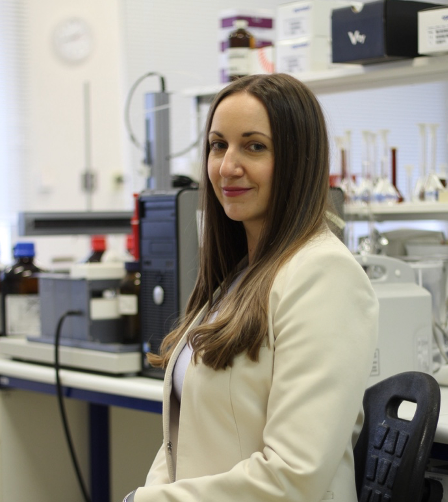
Katarina Marakova of the Department of Pharmaceutical Analysis and Nuclear Pharmacy at Comenius University Bratislava. Photo Credit: © Katarina Marakova.

Can you explain the rationale behind focusing on intact protein analysis using capillary zone electrophoresis-mass spectrometry (CZE–MS) in complex biological matrices?
In the last few decades, protein analysis has shifted from the traditional bottom-up approach into a top-down analysis. It is a powerful approach for studying proteins, including their post-translational modifications (PTMs) and bringing more reliable identification. I started working with intact proteins during my scholarships at Professor Kevin Schug’s laboratory at the University of Texas at Arlington. At that time, I worked on the development of a method for targeting small intact growth factors and cytokines in the biological matrices by liquid chromatography (LC) in hyphenation with triple quadrupole mass spectrometry (2). Even though, LC is a work-horse technique in many applications, capillary electrophoresis (CE) has become an increasingly popular separation technique in proteomic analysis. It has a place in a biopharma industry, and it is an attractive tool also for biomarker discovery. CE is an eco-friendly technique, as shown in our recent paper (3), that provides fast, highly efficient separations and requires ultralow sample and reagent volumes per single analysis (suitable especially for clinical samples). CE separates proteins based on their charge-to-size ratio, leading to high resolution separation and allowing for the identification of closely related proteins.
I have been working with CE during my PhD, my post-doctoral studies, and in our laboratory, we have multiple CE instruments. Therefore, when I returned to my home institution, it was a natural choice for me to focus my research on analyzing intact proteins by CE and CE-MS.
What were the primary challenges associated with targeting intact proteins in complex biological fluids, and how did you address them in your study?
Intact protein analysis in complex biological fluids is associated with multiple challenges. Biological fluids contain a wide range of proteins, including highly abundant serum albumin and immunoglobulins, as well as low-abundance proteins such as cytokines and growth factors. This complexity and heterogeneity make it challenging to detect and quantify specific proteins of interest. Moreover, proteins in biological fluids can be susceptible to degradation or modification during sample handling and storage, what can also affect their detection. When using MS for detection, matrix effects can cause ion suppression or enhancement; therefore, it can affect the accuracy, precision, and reproducibility of protein quantification. When applying CE as a separation technique, you must count with its lower concentration sensitivity compared to LC because of the small injection volume.
Addressing these challenges requires a careful development and optimization of the analytical workflow from sample preparation up to the detection. In our case, we applied CE-MS for targeting intact proteins, and these orthogonal techniques helped to minimize or overcome some problems. CE separates proteins based on their charge, size, and shape, reducing the matrix effects more when compared to techniques such as LC. MS detection offers high sensitivity, allowing for the detection of low-abundance proteins also in complex biological matrices. Finally, a development of an effective sample preparation method is necessary to remove contaminants and enhance assay sensitivity and specificity. To remove potential interferences from the complex biological samples, we used simple microelution solid-phase extraction (mSPE), so the sensitivity of CE was increased by applying the in-capillary electromigration preconcentration method.
Could you describe the key components of your analytical workflow, including the sample pretreatment, separation, and detection steps?
Microelution solid phase extraction (mSPE) was applied in our work for sample preparation. Although mSPE is commonly employed for peptide extraction and purification in proteomics workflows, it is less frequently used for intact protein analysis. However, mSPE can be used to fractionate protein samples based on their physicochemical properties, such as size, charge, or hydrophobicity, thereby enhancing the resolution and sensitivity of subsequent analytical techniques.Although mSPE can be adapted for intact protein analysis, it is important to note that mSPE has a limited capacity and selectivity for intact proteins compared to smaller molecules or peptides. Additionally, the choice of mSPE conditions should be carefully optimized (4) to ensure efficient recovery and purification of intact proteins while minimizing sample loss and nonspecific binding. Here, the key components for optimization process were the selection of stationary phase, multiple sample loading, and elution conditions.
The key components of CE were the separation capillary itself and the buffer system. CE capillaries are typically made of fused silica, which offers thermal stability, and compatibility with a wide range of solvents and buffers used in CE. To minimize protein adsorption and improve separation efficiency, the inner surface of the capillary may be coated with a polymer that reduces nonspecific interactions with proteins. The buffer system used in CE plays a crucial role in controlling the pH, ionic strength, and conductivity of the separation electrolyte. To increase the sample injection and thus also a sensitivity of a method, in-capillary sample preconcentration was applied in our case. There are multiple in-capillary preconcentration methods. However, not all of them are compatible with MS and biological matrices.
The key components of MS for intact protein analysis are the ionization source and the mass analyzer. In our case, we applied a low-resolution triple quadrupole mass spectrometer (QQQ) as a detection step. Although QQQ mass spectrometers are not commonly used for intact protein analysis, they can still be valuable tools for targeted quantification and confirmation of intact proteins in complex samples when used in conjunction with appropriate sample preparation. For development of specific transitions corresponding to target intact proteins, QQQ instruments require careful optimization of collision energy, collision gas pressure, and other ionization conditions to obtain efficient fragmentation and detection.
What criteria were considered in the optimization process for selecting the capillary surface and background electrolyte (BGE)? Why were these factors important?
During the CE optimization process and the selection of capillary inner surface and BGE, the key considerations were their mutual compatibility, compatibility with MS detection and ability to minimize protein adsorption on the capillary wall to prevent sample loss and peak broadening during separation. The BGE composition, including buffer type, pH, and ionic strength, influences the electrophoretic mobility and separation. Typically, the CE-MS coupling requires the use of volatile electrolytes with minimal ionic strength so as not to cause the ion suppression. However, the protein adsorption on the uncoated bare-fused silica capillary can be decreased by using acidic BGEs with higher ionic strength and by adding organic modifiers into the BGE solution. The other option that prevents protein adsorption is using the coated capillaries, where the capillary surface should minimize nonspecific interactions with proteins. In this case, you need to select buffer composition under which the capillary coating will be stable and durable. Overall, the CE parameters were optimized to achieve high separation efficiency, high resolution, sensitive detection, and low protein adsorption.
You mentioned two in-capillary preconcentration methods, transient isotachophoresis (tITP) and dynamic pH junction (DPJ). Can you discuss the advantages and limitations of each method and why tITP was chosen as the optimal technique?
In-capillary preconcentration methods, such as tITP and DPJ, can achieve high sensitivity by concentrating protein analytes in a narrow stacking zone within the capillary leading to enhanced detection limits and improved signal-to-noise ratios. They require minimal sample handling and preparation, reducing the risk of sample loss or contamination and enabling efficient analysis of small-volume biological samples. Both our tested methods, tITP and DPJ, are compatible with MS detection and analyzing biological samples.
tITP concentrate proteins based on their electrophoretic mobilities can be performed rapidly within minutes, making it suitable for high-throughput analysis of intact proteins in clinical or research settings. On the other side, tITP has a limited dynamic range for proteins when high-abundance proteins may saturate the preconcentration zone, limiting the detection of low-abundance proteins. Biological fluids may also contain solutes that can interfere with the preconcentration and separation of proteins in tITP, leading to reduced reproducibility and accuracy of protein analysis. Therefore, other sample pretreatment methods to reduce the complexity of sample matrix before tITP are important.
DPJ can selectively preconcentrate proteins based on their isoelectric point values. DPJ is relatively robust and less sensitive to variations in sample composition and matrix effects, making it suitable for analysis of diverse biological samples. However, DPJ may have limited resolution for closely related protein species. Overlapping peaks and peak broadening may occur, reducing the separation efficiency and peak capacity of DPJ. DPJ may also involve exposure of proteins to extreme pH conditions during the preconcentration process, leading to protein denaturation or degradation.
The choice of the final optimum preconcentration method was based on the obtained preconcentration factor. Optimization of experimental conditions, including sample buffer composition, electric field strength, and sample injection conditions, was crucial to maximize the performance and reliability of these preconcentration methods.
How did offline microscale SPE with hydrophilic lipophilic balance (HLB) sorbent contribute to enhancing the sensitivity of the method? Can you explain the eluate treatment procedures evaluated during SPE?
SPE served as a clean-up step to remove contaminants and matrix components and reduce sample complexity that may suppress protein signals and interfere with CE-MS analysis.
The choice of elution solvent in SPE affects the efficiency and selectivity of protein elution from the SPE sorbent. In our previous work (4), we found out that for reaching the good extraction recoveries, trifluoroacetic acid (TFA) with 75% acetonitrile served as an optimum elution solvent, which was after a simple dilution compatible with direct injection into the LC–MS system. However, the ion suppression caused by TFA was more pronounced when CE-MS analysis was used after SPE. Moreover, both high concentration of acid and organic solvent in the eluate are not compatible with CE separation and disrupt the process of tITP stacking. Therefore, eluate treatments, such as solvent evaporation and freeze drying, were applied to transfer proteins into a more compatible buffer solution for subsequent CE-MS analysis.
What were the extraction recoveries of spiked proteins across different biological matrices, and how did they vary? How were these variations addressed in the method validation process?
The extraction recoveries of spiked proteins across different biological matrices varied widely (21–106%) depending on the protein size, isoelectric point, hydrophobicity, its ionization in MS, and the complexity of the sample matrix. Extraction recoveries of spiked proteins in serum and plasma are generally lower and may be influenced by protein-binding interactions. Urine and saliva samples are less complex; however, extraction recoveries may be affected by pH or salt concentration variations.
To address the matrix effects and variations in extraction recoveries, matrix-matched calibration curves were constructed during validation to ensure accurate quantification of target proteins in biological samples.
Can you summarize the validation results of your optimized method, including assessments of linearity, accuracy, and precision? How do these results support the suitability of your method for proteomic studies in various biological samples?
The linearity of the method was assessed by constructing matrix-matched calibration curves using spiked samples with known concentrations of target proteins that underwent the optimum mSPE pretreatment. Some validation results were not possible to evaluate, as some proteins were migrating together with residual matrix components (probably high-abundance proteins) and in selected ion monitoring mode we were not able to separate them. This could be solved by applying a multiple reaction monitoring (MRM) mode. Other proteins exhibited good linearity in the selected calibration ranges.
Quality control (QC) samples were used to evaluate precision (%RSD) and accuracy (%RE) of the developed method. Acceptable accuracy is typically defined to be within a predefined range (for example, 80–120%). In all biological matrices, the accuracy and precision results met the above-mentioned criteria for many proteins. For some proteins, precision and accuracy data were slightly higher than the required <15 % for %RSD and %RE as stated in validation guidelines. However, those could be improved in future studies by using appropriate internal standards.
Overall, the validation results show the potential to use the developed method for the quantitation of small molecular mass proteins in less complex biological matrices, such as saliva and urine. More complex biological matrices, such as serum and plasma, would need a more selective sample pretreatment protocol based on (immuno)affinity depletion of high abundance proteins or application of more selective MRM mode or high-resolution MS (HRMS).
In what specific applications do you envision this method being particularly useful, such as therapeutic drug monitoring or biomarker research? Can you explain why?
Overall, the developed CE-MS method combined with in-capillary tITP preconcentration and offline mSPE was shown to be suitable for the quantitative determination of intact low molecular mass proteins especially in less complex biological samples (saliva, urine) when the targeted concentration of protein is in hundreds of ng/mL. Therefore, I see the potential to use the developed method for therapeutic drug monitoring of therapeutic peptides and for biomarker research in these less complex biological fluids, especially when the proteins are elevated during a disease.
With some further advances, it can be applied even for a broader range of proteins and applications. However, in more complex biological fluids, such as serum and plasma, trace biomarker analysis, or both, a more selective high-resolution tandem MS approach should be applied to resolve proteins overlapped by the matrix components. In our case, the use of a sheath-liquid in electrospray ionization (ESI) dilutes the sample before entering the MS detector, which also contributes to the sensitivity decrease. Therefore, employing a sheathless interface could result in a more sensitive analysis.
References
(1) Opetova, M.; Tomasovsky, R.; Mikus, P. Maráková, K. Transient Isotachophoresis–Capillary Zone Electrophoresis–Mass Spectrometry Method with Off-line Microscale Solid-phase Extraction Pretreatment for Quantitation of Intact Low Molecular Mass Proteins in Various Biological Fluids. J. Chromatogr. A. 2024, 1718, 464697. DOI: 10.1016/j.chroma.2024.464697
(2) Marakova, K.; Rai, A. J.; Schug, K. A. Effect of Difluoroacetic Acid and Biological Matrices on the Development of a Liquid Chromatography–Triple Quadrupole Mass Spectrometry Method for Determination of Intact Growth Factor Proteins. J. Sep. Sci. 2020, 43 (9–10), 1663–1677. DOI: 10.1002/jssc.201901254
(3) Marakova, K.; Tomasovsky, R.; Opetova, M.; Schug, K. A. Greenness of Proteomic Sample Preparation and Analysis Techniques for Biopharmaceuticals. TrAC Trends Anal. Chem. 2023, 171, 117490. DOI: 10.1016/j.trac.2023.117490
(4) Marakova, K.; Renner, B. J.; Thomas, S. L.; et al. Solid Phase Extraction as Sample Pretreatment Method for Top-down Quantitative Analysis of Low Molecular Weight Proteins from Biological Samples Using Liquid Chromatography - Triple Quadrupole Mass Spectrometry. Anal. Chim. Acta. 2023, 1243, 340801. DOI: 10.1016/j.aca.2023.340801
New Study Uses MSPE with GC–MS to Analyze PFCAs in Water
January 20th 2025Scientists from the China University of Sciences combined magnetic solid-phase extraction (MSPE) with gas chromatography–mass spectrometry (GC–MS) to analyze perfluoro carboxylic acids (PFCAs) in different water environments.
SPE-Based Method for Detecting Harmful Textile Residues
January 14th 2025University of Valencia scientists recently developed a method using solid-phase extraction (SPE) followed by high-performance liquid chromatography coupled to high-resolution mass spectrometry (HPLC–HRMS/MS) for detecting microplastics and other harmful substances in textiles.