Reversed-Phase Analysis of Polysaccharides Using a 2-µm Non-Porous ODS Phase
Polysaccharides are often separated using SEC columns. However, SEC columns do have limitations, specifically: poor column efficiency or peak shape.
Bryan Evans and Itaru Yazawa, Imtakt USA
Polysaccharides are often separated using SEC columns. However, SEC columns do have limitations, specifically: poor column efficiency or peak shape. An alternative to SEC (and CE) for polysaccharide analysis is reversed-phase mode, utilizing Presto FF-C18 (2 μm nonporous ODS).

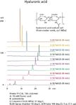
Figure 1: Hyaluronic acid.
Experimental and Results
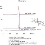
All data was generated with semi-micro HPLC system equipped with ELS detection. Figure 1 shows analysis for hyaluronic acid. Different retention times were obtained by adjusting the beginning or ending organic composition. Figure 2 shows analysis for an endotoxin (a lipopolysaccharide from E. Coli). Figure 3 shows analysis for dextrans (up to 40 MDa). Even though the molecular weight is extremely large, the retention time and peak shape is acceptable for quantification.

Figure 2: Lipopolysaccharide (from E. coli O127).

Figure 3: Dextrans.
Conclusion
Presto FF-C18 (2 μm nonporous ODS) provides an alternative to SEC columns for biopolymer separations.

Imtakt USA
1511Walnut St., Suite 310, Philadelphia, PA 19102
tel. (888) 456-HPLC, (215) 665-8902; fax (501) 646-3497
Website: www.imtaktusa.com; Email: info@imtaktusa.com

SEC-MALS of Antibody Therapeutics—A Robust Method for In-Depth Sample Characterization
June 1st 2022Monoclonal antibodies (mAbs) are effective therapeutics for cancers, auto-immune diseases, viral infections, and other diseases. Recent developments in antibody therapeutics aim to add more specific binding regions (bi- and multi-specificity) to increase their effectiveness and/or to downsize the molecule to the specific binding regions (for example, scFv or Fab fragment) to achieve better penetration of the tissue. As the molecule gets more complex, the possible high and low molecular weight (H/LMW) impurities become more complex, too. In order to accurately analyze the various species, more advanced detection than ultraviolet (UV) is required to characterize a mAb sample.

.png&w=3840&q=75)

.png&w=3840&q=75)


.png&w=3840&q=75)


.png&w=3840&q=75)


