The Potential of Polymer Monolithic Capillary Columns for the LC–MS Analysis of Intact Proteins
LCGC Asia Pacific
Describes the preparation of a reversed-phase monolithic column with an optimized porous structure to separate intact proteins using LC–MS.
LC–MS-based top-down proteomics involves the chromatographic separation and mass-spectrometric characterization of intact proteins without prior enzymatic cleavage into peptides. This article describes the preparation of monolithic reversed-phase (RP) columns with optimized porous structure and their performance separating intact proteins. Peak capacities of around 600 could be achieved within a 2 h gradient. The combination of high-resolution monolith chromatography with high-accuracy timeofflight mass spectrometry (TOF-MS) allowed intact proteins and protein isoforms that differ only in their oxidation state to be distinguished. The article also describes various MS fragmentation techniques for the identification and the more detailed characterization of intact proteins.
The concept of separation media based on monolithic materials can be traced back to the early 1950s, when Nobel Prize Laureates Martin and Synge discussed the benefits of these materials for different applications in chromatography (1). One of the first monolithic polymer gels for liquid chromatography (LC) was developed by Kubín et al. in 1967 (2). Highly swollen hydrogels, prepared in glass tubes from 2-hydroxyethyl methacrylate as the bulk monomer and ethylene dimethacrylate as the cross-linking monomer, were applied for the size-exclusion separations of water-soluble polymers. Rigid macroporous polymer-based monolithic columns were introduced in the early 1990s (3). This type of stationary phase is composed of a single monolithic unit with interconnected microglobules and macropores (flow-through pores), and is typically bonded to the column wall via a covalent bonding to enhance the robustness.

Polymer monoliths are prepared from liquid precursors, that is, the bulk monomer and cross-linker dissolved in the porogen, allowing their in-situ preparation in virtually any format. The separation performance strongly depends on the porous structure of the monolithic material (4). It has been demonstrated that the composition of the polymerization mixture, including the type or ratio of the porogen solvents in the polymerization mixture (5), and the polymerization conditions, such as polymerization temperature and time (6), are key parameters that need to be controlled precisely. The surface chemistry and therefore selectivity can be tuned by incorporating 'functional' monomers in the polymer backbone (7). As a result of the absence of mesopores (stagnant zones inside polymer microglobules), the mass-transfer contribution to total band broadening is greatly reduced (8).
The success of polymer monolithic columns for the reversedphase gradient-elution separation of peptides in a LC–MS bottom-up proteomics approach has been demonstrated on several occasions (9–11). Karger et al. explored the use of 20 µm i.d. monolithic columns and established an HPLC–ESI–MS method yielding low-attomol detection sensitivity (12). Recently, we demonstrated a LC–MS–MS separation of a tryptic E. coli digest on a 1 m monolithic column yielding a peak capacity in excess of 1000 (13).
LC–MS-based top-down proteomics is based on the chromatographic separation of intact proteins followed by their mass spectrometric elucidation. Single-stage MS provides information on the molar mass of intact proteins. The mass alone can give information about chemical or post-translational modifications of proteins, provided that the identity of the protein is already known. Tandem mass spectrometry MSn can provide structural information and is essential for the identification of unknown proteins. In MSn the selected precursor ions are fragmented in the gas phase using neutral gas molecules for collision-induced dissociation (CID) (14) and higher-energy collisional dissociation (HCD) (15); electrons for electron capture dissociation (ECD) (16); or radical anions for electron transfer dissociation (ETD) (17). In top-down proteomics high-resolution separation technology is of vital importance to address two bottlenecks: ion suppression and the spectral complexity.
Here, the potential of capillary polymer monolithic columns is discussed for the separation of intact proteins, including protein isoforms. The effects of column properties (length and morphology) and gradient conditions on the separation performance were evaluated. The potential to achieve highresolution protein separation is demonstrated with the liquid chromatography time-of-flight mass spectrometry (LC–TOF-MS) analysis of a mixture containing 48 intact human proteins. In addition, the application of complementary fragmentation techniques (CID, HCD and ETD) is demonstrated for protein identification using a hybrid ion trap-orbitrap mass spectrometer.
Experimental
Chemicals and materials: Divinylbenzene (DVB, 65%) and styrene (S, > 99%) were purchased from Merck (Hohenbrunn, Germany). Sodium hydroxide pellets were obtained from Merck (Overijse, Belgium). Acetonitrile (ACN, HPLC supragradient quality), formic acid (FA, ULC/MS quality), tetrahydrofuran (THF, > 99.9 %), aluminium oxide (Al2O3), toluene (99.8 %), 3-(trimethoxysilyl)propyl methacrylate (98 %), methanol, 1-decanol (> 95.0 %, GC quality), trifluoroacetic acid (TFA, ULC/MS quality), carbonic anhydrase from bovine erythrocytes, ribonuclease A from bovine pancreas, lysozyme from gallus egg white, myoglobin from equine heart, trypsinogen from bovine pancreas and the "Universal Proteomics Standard 1" containing 48 human source or human sequence recombinant proteins (ABRF mixture) were purchased from Sigma-Aldrich (Bornem, Belgium).
DVB and S monomers were purified by passing them over activated basic alumina. Water was purified in-house using a Millipore Simplicity system (Millipore, Bedford, MA, USA). Fused-silica capillary tubing (200 µm i.d. × 365 µm o.d.) was purchased from Aventes (Eerbeek, The Netherlands).
Instrumentation: HPLC experiments were performed using an UltiMate 3000 Proteomics MDLC system (Dionex Corporation, Germany) equipped with a membrane degasser, ×2 dual-gradient pump, a thermostatted flow manager and well-plate sampler applying a linear aqueous ACN gradient. For LC–TOF-MS analysis of intact proteins the HPLC system was coupled on-line with a maXis ultra-high-resolution qTOF mass spectrometer via a nano-electrospray interface (Bruker Daltonics, Bremen, Germany) equipped with a Proxeon steel emitter. Spectra were obtained in positive ion (PI) mode. LC–TOF-MS analysis of intact protein was performed on a 250 mm × 0.2 mm i.d. PepSwift monolithic capillary column purchased at Dionex Benelux.
An Orbitrap Elite hybrid mass spectrometer (Thermo Fisher Scientific, Bremen, Germany) was used for MS–MS fragmentation experiments. Protein solutions were infused directly into the mass spectrometer. A solution of beta2microglobulin (0.5 µg/µL in 1:1 water/acetontrile containing 0.1% formic acid) was infused into the Orbitrap Elite mass spectrometer (Thermo Scientific) at 5 µL/min. The source voltage was set to 5 kV and source temperature to 275 °C. The AGC settings was 1E6 and 500 ms for MS and 5E4 and 250 ms for MS/MS. Five to 10 microscans were averaged for MS and MS–MS spectra.
Scanning electron micrographs (SEMs) were obtained using a field emission scanning electron microscopy (FE-SEM) (Jeol JSM-7000F FE-SEM, Jeol Ltd., Tokyo, Japan) after sputtering a 20 µm coating on the monoliths with a Cressington 208 HR sputter coater.
Preparation of monolithic columns: To demonstrate the effect of porogen composition of morphology, monolithic columns were prepared in-situ in 200 µm i.d. fused-silica capillaries from styrene and divinylbenzene precursors through a thermally initiated free-radical copolymerization reaction. Prior to the polymerization reaction, surface modification of the inner wall of fused-silica capillaries was performed with 3-(trimethoxysilyl)propyl methacrylate following an approach described by Svec (18). Polymerization mixtures comprising 40 w% monomers (20% S as bulk monomer and 20% DVB as cross-linker), 60 w% porogen (THF and decanol),and initiator (AIBN, 1 w% with respect to monomers) were degassed in an ultrasonic bath for 10 min. Surface-modified capillaries were filled with the polymerization mixture and then sealed with a septum. After polymerization at 70 °C overnight in a water bath, the columns were flushed with methanol and mobile phase.
LC–TOF-MS analysis of intact proteins was performed on a 250 mm × 0.2 mm i.d. PepSwift monolithic capillary columns (Dionex Benelux, Amsterdam, The Netherlands).
Results and Discussion
Control of monolithic structure and effect on gradient performance: Figure 1 shows scanning electron micrographs of the monolithic materials prepared. The close-up [Figure 1(b)] of the monolithic structure depicted in Figure 1(a) clearly shows the typical interconnected polymer microglobules and macropores in the polymeric structure. As a result of the absence of mesopores (stagnant zones in microglobules), polymer monolithic materials are conceptually very well-suited to perform large-molecule gradient separations because mass transfer is mainly driven by convection, rather than by diffusion. The monolith is covalently bonded to the fusedsilica capillary surface via a 3-(trimethoxysilyl)propyl methacrylate spacer to prevent channeling effects of analytes between the monolith and the wall.
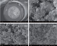
Figure 1
The porous structure of PS-DVB monoliths was systematically varied by changing the porogen ratio (THF/decanol) to maximize the peak capacity. With a decreasing amount of THF in the polymerization mixture, an increase in both the macropore and microglobule size can be observed (Figure 1). These findings can readily be explained by the solvating properties of the porogens: while THF is a thermodynamically "good" solvent for the polymer, decanol constitutes a "poor" solvent. Decreasing the amount of THF (thereby increasing the concentration of "poor" solvent) results in an earlier phase separation and results in the formation of a porous monolithic structure containing large macropores. Since the polymerization reaction is mainly continued within the nuclei, the size of the polymer microglobules increase. The resulting polymer monolith will accordingly exist of large polymer microglobules with large voids (macropores). THF is a "good" solvent for both monomers and polymers and increasing the THF content in the polymerization mixture means the phase separation is postponed. As a consequence, the monomer concentration in the nuclei is smaller, leading to the formation of a larger number of microglobules that are smaller in size [see the monolithic structure depicted in Figure 1(d)].
Columns of different length and morphology were evaluated in gradient LC mode by separating a standard mixture of proteins (ribonuclease A, lysozyme, trypsinogen, myoglobin and carbonic anhydrase) and varying the gradient duration. The peak capacity (nc) was determined by averaging the 4σ peak width (W) and applying Equation 1:

where tG is the gradient time, L is the column length, t0 is the column hold-up time, ke the retention factor of the analyte at the moment of elution and H is the plate height. Figure 2 shows the effect of gradient time on peak capacity (a) and peak-production rate (peak capacity per unit time) (b) operating the columns at a flow rate of 2 µL/min. While longer gradient times and shallower gradients yield higher peak capacities, the peak capacity tends to reach a limit at longer gradient duration due to a linear increase in W. However, with increasing gradient time, the peak-production rate dramatically decreases. Monoliths with smaller macropores and microglobules perform better in terms of peak capacity and in peak-production rate. Also, the optimum gradient time to achieve the maximum peakproduction rate shifts to slightly higher values [Figure 2(b)]. The longer monolithic column performs better in terms of peak capacity and peak-production rate when applying gradient times > 50 min, which is far from the optimal gradient duration required to reach the maximum peakproduction rate. However, this may be alleviated with the development of future monolithic nanomaterials that can be operated at ultra-high pressures.
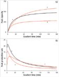
Figure 2
LC–TOF-MS analysis of intact proteins
The potential of long monolithic columns for highly demanding separations is further demonstrated by a top-down LC–TOF-MS analysis of a complex mixture of intact proteins on a 250 mm monolithic column applying a 2 h gradient (Figure 3). The protein mixture contains 48 intact human proteins and the sample complexity is further increased by the presence of protein isoforms arising from co- and post-translational modifications, including oxidation, methylation and glycosylation (19, 20). The maximum peak capacity was around 600. The threedimensional representation of the LC–TOF-MS analysis is shown in Figure 3(b), depicting the retention time (x-axis), the mass-to-charge ratio (m/z) (y-axis) and the signal intensity (z-axis). Figure 3(c) shows the charge envelope of a single protein with charge states ranging between 6+ and 10+ eluting between 46–48 minutes. After deconvolution of the mass spectrum the relative molecular weight (Mr) was determined to be 8,381.4 Da, which corresponds to the theoretical molecular weight of interleukin-8 (Mr = 8,381 Da). Figure 3(d) shows a zoom spectrum of the isotopically resolved 8+ ion of this polypeptide (resolution ≈ 30,000 FWHM).

Figure 3
Figure 4 clearly shows the high information content of the three-dimensional representation of proteins eluting between 88 min and 95 min. Whereas the TIC representation would only show co-eluting proteins, the three-dimensional representation allows for the deconvolution of co-eluting proteins, based on differences in m/z values. Protein identification, as shown in Table 1, was based on matching of experimental Mr values with theoretical masses obtained from the list of known proteins present in the 48 protein standard. The six charge envelopes that were baseline separated were consistent with their allocation to isoforms of peptidylprolyl cistrans isomerase A. The mass difference of 16.0 Da could be indicative of the oxidation of methionine or cysteine residues.
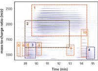
Figure 4
The three-dimensional representation in Figure 5(a) shows highly similar charge envelopes, indicative of the elution of protein isoforms, which are baseline separated. The average molecular weight of the most intense signal (signal 1) was determined to be 11,729 Da. The experimentally determined Mr values coincide well with the expected mass of beta-2-microglobulin (Mr = 11,731 Da). The mass difference of 2 Da can be explained by the formation of a disulfide bridge, which is a documented post-translational modification for this particular protein. The mass difference of 16 Da between peaks 1 and 2 could indicate protein oxidation. This post-translational modification makes the protein more polar and results in a decrease in retention time because of the reversed-phase (RP) character of the stationary phase.

Table 1
Application of complementary MS–MS fragmentation techniques: MSn experiments on the intact protein can add further confirmation to protein sequence and to the characterization of post-translational modification(s). MS–MS experiments were performed on beta-2-microglobulin (~12 kDa) using an Orbitrap Elite hybrid mass spectrometer which is capable of 240,000 resolution (at m/z = 400). Ion trap CID, HCD and ETD were performed on the +12 charge state of beta-2-micoglobulin and the HCD and ETD MS–MS spectra are shown in Figures 6(a) and (b), respectively.

Figure 5
The multiply-charged ions in the ion trap CID and HCD spectra were assigned to b- and y-fragment ions, and sequence coverage with HCD fragmentation was found to be higher compared to ion trap CID. The ETD spectrum in Figure 6(b) revealed a homologous series of c- and z fragment ions. The sequence coverage of applying ion trap CID, HCD and ETD fragmentation is demonstrated in Figure 6(c). Applying complementary fragmentation techniques provided more reliable and detailed aminoacid sequence information. It is important to note that beta-2-microglobulin contains a disulfide bridge and fragmentation was suppressed in the disulfide bonded loop (between amino acid 25 and 80). More structural information may be obtained after reducing the disulfide bridges before the LC–MS–MS experiment (21).

Figure 6
Conclusions
The separation performance of capillary polymer monolithic columns that differ in porous structure and column length was evaluated and the gradient duration was optimized for the LC–MS analysis of intact proteins. The separation performance of the columns was assessed in terms of peak capacity and peak-production rate. Peak capacity can be interpreted as a direct measure for separation power, while peak-production rate can be used as a measure for effectiveness. Longer gradient times and shallower gradients yielded the highest peak capacities. However, with increasing gradient time, the peak-production rate decreased.
The analysis of intact proteins using LC–TOF-MS allowed the tentative identification of proteins based upon their molecular weight. As demonstrated, this may be more confidently achieved by the application of MS/MS fragmentation techniques. The application of highresolution monolith chromatography in combination with complementary fragmentation techniques such as CID, ETD and HCD provides a unique platform to analyse proteins, including isoforms. Reduction of disulfide brides prior to LC–MS–MS analysis might be beneficial to obtain higher sequence coverage.
Acknowledgements
This work was supported by a grant from the Research Foundation Flanders (G.0919.09) and is gratefully acknowledged. Bert Wouters acknowledges the financial support Institute for the Promotion of Innovation through Science and Technology in Flanders (IWT-Flanders). NEPAF is a CELS project funded by ONE NorthEast and partly financed by the European Regional Development Fund (ERDF). Franck van Veen (Dionex Benelux, Amsterdam, The Netherlands) is acknowledged for donation of the PepSwift monolithic columns.
Bert Wouters obtained his Masters degree in Applied Engineering (Chemistry) in 2007 from the Artesis College University of Antwerp, and his Masters degree in Biomolecular Science in 2010 from the Vrije Universiteit Brussel in Brussel. He is a PhD student at the Department of Chemical Engineering at the Vrije Universiteit Brussel and focuses on the development of novel spatial multi-dimensional chromatography.
Axel Vaast is a PhD student at the Department of Chemical Engineering at the Vrije Universiteit Brussel and focuses on the development and characterization of novel polymer monolithic structures for application in chromatography. He obtained a Masters degree in Bioengineering, medical biotechnology (Cellular & Genetic Biotechnology) with great distinction in 2009 from the Vrije Universiteit Brussel.
Achim Treumann is Scientific Manager at NEPAF and is a visiting Research Professor at Northumbria University. After his post-doctoral research at the Fujita Health University in Toyoake, he was appointed Director of Mass Spectrometry in the Department of Clinical Pharmacology at the Royal College of Surgeons (RCSI), Dublin. He has performed extensive work in the application of NMR and mass spectrometry to solve the structure of lipoarabinomannan at both the University of St Andrews and Leeds University. Achim's key areas of expertise include analytical biochemistry, protein chemistry, liquid chromatography, mass spectrometry and bioinformatics.
Mario Ursem is co-founder of LC Packings and is currently Managing Director of Dionex Benelux (Amsterdam, The Netherlands). His interests include column technology for nano- and bio-LC and LC–MS application development.
Jenny TC Ho received her PhD degree in Chemistry (specializing in biological mass spectrometry) from the University of Manchester, United Kingdom. After postdoctoral research positions at the National Institutes of Health and California Institute of Technology she joined Thermo Fisher Scientific as a Proteomics Applications Specialist working on the Orbitrap mass analyser.
Martin Hornshaw is the mass spectrometry Marketing Manager for Thermo Fisher Scientific in Europe. He has over 20 years of experience working with biological mass spectrometry.
Herman Terryn is a professor in the Materials and Chemistry department of Vrije Universiteit Brussel and is responsible for the surface analysis facilities. He is also based part-time as a professor at TUDelft where he is cluster leader of the research on durability of materials within M2i.
Marc Raes is operator of the FESEM-equipment.
Sebastiaan Eeltink received his PhD degree in Chemistry (specializing in Analytical Chemistry) in 2005 from the University of Amsterdam, The Netherlands. After his postdoctoral research at the University of California in Berkeley, he joined Dionex and conducted research on packed and monolith column technology for ultrahighpressure LC, two-dimensional LC and nano-LC. In 2009, the National Fund for Scientific Research (FWO, Belgium) awarded him an Odysseus grant. He currently holds a position as assistant professor at the Department of Chemical Engineering at the Vrije Universiteit Brussel.
References
1. D.L. Mould et al., Electrokin. Ultrafiltr, 58(4), 571–585 (1954).
2. M. Kubin et al., Collect. Czech. Chem. Commun., 32(11), 3881–3887 (1967).
3. F. Svec et al., Anal. Chem., 64(7), 820–822 (1992).
4. J. Mohr et al., Anal. BioAnal. Chem., 400(8), 2391–2404 (2011).
5. S. Eeltink et al., J. Sep. Sci., 30(3), 407–413 (2007).
6. E.C. Peters et al., Macromolecules, 32(19), 6377–6379 (1999).
7. C. Ericson et al., Anal. Chem., 72(1), 81–87 (2000).
8. M. Petro et al., Anal. Chem., 68(3), 315–321 (1996).
9. J. Zhang et al., J. Chromatogr. A, 1154(1–2), 295–307 (2007).
10. X. Huang et al., Anal. Chem., 74(10), 2336–2344 (2002).
11. A. Tholey et al., Anal. Chem., 77(14), 4618–4625 (2005).
12. A.R. Ivanov et al., Anal. Chem., 75(20), 5306–5316 (2003).
13. S. Eeltink et al., J. Chromatogr. A, 1217(43), 6610–6615 (2010).
14. C.A. Redman et al., Glycoconjugate J., 11(3), 187–193 (1994).
15. Y. Zhang et al., J. Am. Soc. Mass Spectrom., 20(8), 1425–1434 (2009).
16. K.F. Medzihradszky et al., Mol. Cell. Prot., 3(9), 872–886 (2004).
17. J.E.P. Syka et al., P. Natl. Acad. Sci. USA, 101(26), 9528–9533 (2004).
18. T. Rohr et al., Macromol., 36(5), 1677–1684 (2003).
19. S. Eeltink et al., J. Chromatogr. A, 1218(32), 5504–5511 (2011).
20. L. Bianco et al., J. Proteome Res., 8(4), 1782–1791 (2009).
21. J.L. Stephenson et al., Rapid Commun. Mass Spectrom., 13(20), 2040–2048 (1999).

