Glycosylation Analysis Through Released N-Glycan Workflows
Post-translational modifications are potential critical quality attributes (pCQAs) routinely assessed in biotherapeutic development. Glycosylation is one of the most important attributes to assess because it affects protein function as well as antigen receptor binding. N-glycosylation of asparagine residues is the most common pCQA assessed during monoclonal antibody (mAb) therapeutic development. There are a few protocols to assess and quantitate N-glycans, but the most common approach is through an enzymatic release and labelling procedure, followed by separation and detection. This article demonstrates the method development considerations for sample preparation and chromatographic analysis of N-glycans of therapeutic mAbs.
sakurra/stock.adobe.com
Post-translational modifications are potential critical quality attributes (pCQAs) routinely assessed in biotherapeutic development. Glycosylation is one of the most important attributes to assess because it affects protein function as well as antigen receptor binding. N-glycosylation of asparagine residues is the most common pCQA assessed during monoclonal antibody (mAb) therapeutic development. There are a few protocols to assess and quantitate N-glycans, but the most common approach is through an enzymatic release and labelling procedure, followed by separation and detection. This article demonstrates the method development considerations for sample preparation and chromatographic analysis of N-glycans of therapeutic mAbs.
Glycosylation events are fundamental in biology and necessary for a plethora of biological processes. The most recent example is evident in the advent of the coronavirus (COVID-19) pandemic, where glycosylation has been predicted to contain “unique N- and O-linked glycosylation sites of spike glycoprotein that distinguish it from the SARS and underlines shielding and camouflage of COVID-19 from the host defence system” (1). In biotherapeutic development, glycosylation has many implications in safety, efficacy, and immunogenicity. In monoclonal antibody (mAb) development, potency depends on biological effector functions and glycosylation plays an important role in this regard (2). Moreover, the pharmacokinetics (PK) of mAbs is significantly altered based on the glycosylation pattern (3). Arguably, the two most important N-glycan types are high mannose forms, such as Man5 and Man6, and sialylation because their impact in efficacy and PK profile is established. As such, researchers must consider glycosylation as one of the more important post-translational modifications (PTMs) to monitor because of these profound biological effects. Therefore, many methods have been developed for quantitation of glycans of biotherapeutics.
Structurally, in the most common type of mAb therapeutic, immunoglobulin G 1 (IgG1), glycosylation occurs covalently at the CH2 domain at asparagine 297 (Asn-297) (4). Because this glycosylation location is inherent to IgG1 mAbs, site-specific cleavage of the Asn-297 residue can reveal the glycans attached to the mAb. The structure of these N-linked glycans can be assessed by intact analysis methods (5,6), however, N-linked glycan by release is the most accurate way to characterise the heterogeneity of glycosylation because lower level glycoforms cannot be detected by glycopeptide nor at the intact level (5). The enzyme PNGase F selectively cleaves N-glycans at the Asn-297 residue, which then must be labelled with an appropriate dye and subsequently separated by hydrophilic interaction chromatography (HILIC) for quantitation. This article demonstrates the released N-glycan analysis of proteins through different labelling techniques and focuses
on high mannose and sialylation detection. Finally, an analysis of biosimilars shows the importance of N-glycan analysis during therapeutic development.
Materials and Methods
Reagents/Chemicals: Alpha-1 acid glycoprotein (AGP) sample derived from human plasma and 2-aminobenzamide (2-AB) were obtained from Sigma-Aldrich. Infliximab and biosimilars were obtained from Myoderm. 2-AB-labelled glycan libraries, PNGase F, and Gly-X N-Glycan Rapid Release and InstantPC kit were obtained from ProZyme (now part of Agilent). GlycoWorks RapiFluor Labelling Module was obtained from Waters. The bioZen N-Glycan Clean-Up plate and bioZen 2.6-µm Glycan column were from Phenomenex.
Experimental Conditions: To prepare infliximab and biosimilars for chromatographic analysis, the Gly-X and InstantPC kit was utilized. The protein was first denatured with the denaturation reagent provided and incubated at 90 °C for 3 min. Subsequent enzymatic digestion with PNGase F at 50 °C for 5 min provided the released glycans. Immediate labelling with InstantPC dye for 1 min at 50 °C followed by clean-up through a bioZen N-Glycan Clean-Up Plate provided the labelled N-glycans. 1 μL of the elution from the glycan cleanup plate was injected on a bioZen 2.6 μm Glycan column.
AGP was reconstituted to 2 mg/mL in pure water. Each sample was denatured by adding 6 µL of surfactant and heating to 90 °C for 3 min. 1.2 µL of PNGase F was then added and samples were incubated for 5 min at 50 °C. Released glycans were labelled with 12 µL of labelling reagent solution for each sample and incubated for 5 min at room temperature.
Results and Discussion
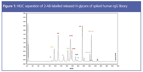
The most traditional and adopted protocol for N-glycan analysis deploys a 2-aminobenzamide (2-AB) label to the released glycans (7). This reductive amination process is robust and provides sufficiently fluorescent materials for analysis by a fluorescence detector (FLD). To demonstrate this technique, the prelabelled human IgG library was assessed. The 2-AB human IgG library was then subsequently spiked with 2-AB-labelled Man5 and Man6 to give a better representation of a recombinantly expressed protein which may have immature glycans. Samples were then analyzed using a HILIC LC column (Figure 1). Notably, there is baseline separation of Man5 and Man6 from other glycan species, which allows quantitative or qualitative assessment of these pCQAs pending the analytical goal.
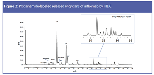
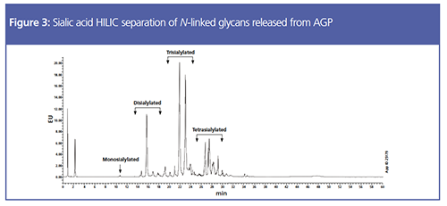
An alternative approach to the labelling step is to use a procainamide label via a carbamate linkage, which has better sensitivity by FLD. An infliximab biosimilar was assessed using this labelling method, but still under HILIC separation (Figure 2). Notably, the Man5 species elutes much closer to G0 under this protocol indicating that the 2-AB labelling method is more efficient at this specific separation. Another aspect of this data to observe is the sialic acid region of the chromatogram. As mentioned earlier, sialylation affects the PK results, so it is important to obtain sufficient separation in this region of the chromatographic profile. To clearly demonstrate the sialic acid separation, N-linked glycans released from serum-derived AGP were assessed (Figure 3). This model protein is a suitable surrogate for a complex, highly sialylated glycoprotein with both neutral and sialylated glycans with multiple antennae. In this case, a quinolinyl label was employed which is beneficial for sensitive mass spectrometry (MS) analysis (8). The chromatogram in Figure 3 was obtained by increasing the ammonium formate in the mobile phase to 250 mM, which was necessary to partition the sialylated species.
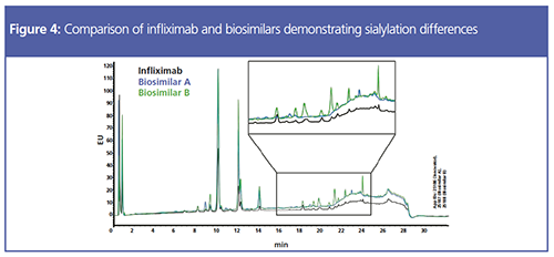
Finally, infliximab and biosimilars were analyzed to determine glycosylation differences in these commercially-available mAbs (Figure 4). The results from this assessment indicate relatively similar levels of glycosylation of the neutral species, but significant variation in sialylation (zoomed region). Clearly, because the biosimilars will be generated from different cell lines and fermentation processes, glycosylation PTMs are expected. As these are all commercially-available therapeutics, these specific glycosylation differences are likely to have minimal therapeutic impact.
Conclusion
Monoclonal antibody therapeutics are complex mixtures of protein variants with glycosylation being a PTM of critical therapeutic importance. The most common approach to assess these glycans is through enzymatic release of N-glycans using PNGase F followed by labelling with an appropriate dye. This article demonstrates the use of three different fluorescent labels followed by HILIC separation, all of which are efficient at N-glycan quantitation. Ensuring a robust, transferrable, and reproducible method is essential for researchers that characterize and monitor CQAs. Optimization of the protocol may be necessary pending the CQA identified through orthogonal research as outlined by the sialic acid assessment of AGP. Finally, the applications presented here provide various labelling protocols for comparable released N-glycan analysis by HILIC separation.
References
- N. Vankadari, and J.A. Wilce, Emerging Microbes & Infections9(1), 601–604 (2020).
- R. Jefferis, Biotechnology Progress 21(1), 11–16 (2005).
- L. Liu, J. Pharmaceutical Sciences104(6), 1866–1884 (2015).
- F. Higel, A. Seidl, F. Sorgel, and W. Fiess, European Journal of Pharmaceutics and Biopharmaceutics100, 94–100 (2016).
- L. Zhang, S. Luo, and B. Zhang, mAbs8, 205–215 (2016).
- C.M. Woo, A.T. Iavarone, D.R. Spiciarich, K.K. Palaniappan, and C.R. Bertozzi, Nature Methods12, 561–567 (2015).
- L.R. Ruhaak, G. Zauner, C. Huhn, C. Bruggink, A.M. Deelder, and M. Wuhrer, Analytical and Bioanalytical Chemistry397, 3457–3481 (2010).
- T. Keser, T. PaviÄ, G. Lauc, and O. Gornik, Frontiers in Chemistry6, 324 (2018).
Christina Malinao received her B.S. in neuroscience and biology from the University of California, Riverside (USA). She spent some time at Schering Plough performing biomarker characterization, and several years at Agensys performing antibody and antibody–drug conjugate characterization. She joined Phenomenex as a scientist in the Phenologix application laboratory performing method development. Christina currently works at Tae Life Sciences.
Brian Rivera is the Senior Product Manager – Biologics at Phenomenex. He has worked at Phenomenex for nine years, holding other positions including technical support and sales. Prior to joining Phenomenex, Brian worked within the biotechnology industry, including positions focused on protein purification, analytical method development, and in-process analytical support. Brian has a bachelor’s degree from the University of California, Davis, USA.
Chad Eichman is the Global Business Unit Manager – Biopharmaceuticals at Phenomenex, where he develops and leads the strategic plans for Phenomenex’s biopharmaceutical business. He received his B.S. in chemistry from the University of Wisconsin–Madison (USA) and his Ph.D. in organic chemistry from The Ohio State University (USA). After a postdoctoral appointment at Northwestern University (USA) and an assistant professorship at Loyola University Chicago (USA), Chad joined Phenomenex in 2017.
E-mail:ChadE@phenomenex.comWebsite:www. phenomenex.com/bioZen
Accelerating Monoclonal Antibody Quality Control: The Role of LC–MS in Upstream Bioprocessing
This study highlights the promising potential of LC–MS as a powerful tool for mAb quality control within the context of upstream processing.
Common Challenges in Nitrosamine Analysis: An LCGC International Peer Exchange
April 15th 2025A recent roundtable discussion featuring Aloka Srinivasan of Raaha, Mayank Bhanti of the United States Pharmacopeia (USP), and Amber Burch of Purisys discussed the challenges surrounding nitrosamine analysis in pharmaceuticals.

.png&w=3840&q=75)

.png&w=3840&q=75)


.png&w=3840&q=75)


.png&w=3840&q=75)


