Charge Detection Mass Spectrometry: What’s the “Big” Deal?
Over the last several years, the landscape of the biopharmaceutical industry has begun to change. Monoclonal antibodies (mAbs) still dominate the pipelines in the biopharmaceutical industry; however, more complex molecules, such as antibody–drug conjugates (ADC), are becoming more predominant. In addition to the complexity of ADCs, one of the fastest growing classes of biopharmaceuticals is cell and gene therapies. Gene therapies, for example, use large viral vectors, such as adeno-associated viruses (AAV), as the preferred vector for performing gene therapy. With the complexity of biopharmaceuticals increasing, especially in their size, new and innovative approaches are needed to address and characterize these molecules. This month’s column reviews how charge detection mass spectrometry (CDMS) is being used to characterize some of these larger more complex molecules in the biopharmaceutical industry.
Biopharmaceuticals are products that are specifically made by living cells or biotechnological processes. By the nature of these products being manufactured in living cells can lead to variability in the product produced, that is the “process is the product” (3). In addition, compared to synthetic drugs, biopharmaceuticals are orders of magnitude larger and more complex because they often contain post-translation modifications such as glycosylation and can form higher-order structures (oligomers). These post-translation modifications are largely why biopharmaceutical products are so complex and a diverse set of analytical tools is needed for their characterization (3).
The biopharmaceutical industry is a billion-dollar industry that is largely dominated by monoclonal antibodies (mAbs). The first mAb was registered in 1986, and the 1990s saw the rapid evolution of biopharmaceutical development and products entering the market. In 2002, the first fully humanized mAb was approved by the U.S. Food and Drug Administration (FDA) (3). Thus, the biopharmaceutical industry is still in its infancy and new, more complex products are in development and will likely dominant the market in the future. For example, several bispecific antibodies are in clinical trials. A bispecific antibody is an artificial protein composed of fragments from two different mAbs, which allows them to bind to different antigens. The bispecific nature of these antibody products makes them promising therapies for certain types of cancer. Another promising new modality is antibody–drug conjugates (ADCs), which are bioconjugated products. In an ADC, a mAb is conjugated with a potent drug, and the mAb targets a specific antigen and delivers the drug to the target. ADCs have proven to be effective at specifically targeting and delivering anti-cancer drugs. Although these are only a few examples of the more complex biopharmaceuticals in development, there are many others such as glycoengineered mAbs, recombinant protein therapeutics, vaccines, and gene therapies (3).
The development and manufacture of these new, more complex biopharmaceuticals drive the need for new analytical technologies to characterize them. Arguably, the most common analytical tool for characterizing biologics is liquid chromatography–mass spectrometry (LC–MS). However, the most common forms of LC–MS have limitations when characterizing large macromolecules (4). Thus, in this column, we discuss the potential for charge detection MS (CDMS) as an analytical tool for characterizing large, complex, and heterogenous biopharmaceuticals.
Native Mass Spectrometry (Native MS)
Before discussing CDMS, we should briefly discuss native MS, which is perhaps the most commonly used technique to characterize the intact mass of large, complex biomolecules in the megadalton range. As with all MS experiments, sample preparation is of utmost importance, which is also true for native MS. Proteins or protein complexes are isolated, purified, and then often diluted in a solvent such as ammonium acetate for the efficient spraying through the electrospray ionization (ESI) emitter into a MS instrument (5).
When a quadrupole time-of-flight instrument (QTOF) is used in native MS, the ions of the intact molecules pass through the quadrupole and through the collision cell, which is turned off.The parameters of the quadrupole are adjusted to promote the transmission of high mass-to-charge (m/z) ratio ions. For example, in the quadrupole, the pressure of the pumping stages is increased and the frequency is decreased, allowing the transmission of ions in the thousands of m/z range (5).
These modifications allow for the characterization of protein–protein and protein–ligand complexes. However, there are limitations to this approach. Namely, the molecular mass of the macromolecule can only be determined if the mass and charge state can be resolved for multiple charge states of the molecule. In addition, there is often poor desolvation and broadened mass distributions, which result in overlapping signals between consecutive charge states. These limitations can lead to inaccurate mass assignments preventing the characterization of large heterogeneous proteins or complexes, such as highly glycosylated proteins, aggregates, and genome packed viruses like adeno-associated virus (AAVs) (4).
Charge Detection Mass Spectrometry (CDMS)
One approach to overcome the convolution of ions, as mentioned above, is to employ single particle, or ion, detection. When single particle detection is paired with the identification of the charge of the ion, we are left with single-particle MS (sp-MS) measurements. There are several techniques for sp-MS detection, one being CDMS, which has gained momentum over the last few years in analyzing complex biopharmaceuticals (4). As a matter of fact, in October 2021 at the American Association for Mass Spectrometry (ASMS) annual meeting in Philadelphia, TrueMass presented the first commercial CDMS (6,7). Other companies, such as Megadalton Solutions (8), offer fee-for-service analysis of protein assemblies, aggregates, AAV, and vaccines.
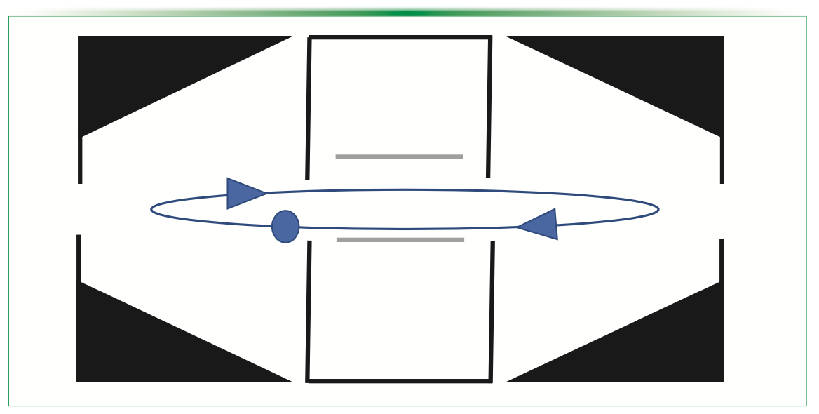
In CDMS, the masses of individual ions are determined by the simultaneous measurement of the m/z ratio of each ion and the charge itself (2). In a CDMS experiment, the m/z of the individual ions and charge are measured, whereas in traditional MS experiments only the m/z ratio is measured, and the charge is inferred from the charge state envelope or distribution. The charge in a CDMS experiment is measure by passing the ions through a conducting cylinder where the charge of the ion is induced on the cylinder and detected. The cylinder is often inside an electrostatic linear ion trap (ELIT) instrument, where ions oscillate back and forth. Therefore, the oscillation frequency gives the m/z and the charge is determined by the magnitude. Historically, the resolving power has been modest, but in recent years and likely in the future, the resolving power will continue to improve (2).
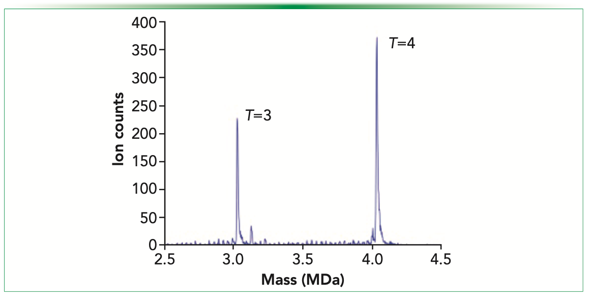
For example, in 2020, Todd and others showed an order of magnitude increase in the resolving power of CDMS by combining dynamic calibration with improvements in an ELIT instrument (2). Briefly, when considering using an ELIT instrument in CDMS, the conducting cylinder is placed in an ELIT instrument, allowing the ions to oscillate in the cylinder many times as opposed to the one time in CDMS experiments where the conducting cylinder is not placed in an ELIT instrument (Figure 1). In dynamic calibration, the charge-sensitive amplifier is calibrated during the measurement by placing a small antenna in the outer casing of the ELIT instrument (9). This approach of combining dynamic calibration and ELIT improvements, leading to increased resolving power, was benchmarked by the analysis of trapped intermediates in the assembly of the Hepatitis B virus (2), thus showing its applicability to not only large molecule detection but in analysis of heterogeneous samples (Figure 2).
FIGURE 1: Schematic of charge detection mass spectrometry (CDMS). Adapted from JASMS (1).

FIGURE 2: A representative example of CDMS spectra. Higher resolving power CDMS mass spectrum measured for HBV Cp149 assembly reaction. There are prominent peaks attributable to the icosahedral T = 3 and T = 4 capsids. The bin size is 2.5 kDa. Reproduced with permission from Analytical Chemistry (2).

In another study in 2020 by Worner and others, they showed the use of an orbital trap MS in CDMS analysis. In this study, the authors were able to characterize a complex mixture of IgM oligomers and co-occurring empty and genome loaded AAV8 particles. Of particular note in characterizing these molecules, the authors showed the application of CDMS to analyzing heterogenous complexes where conventional native MS cannot be used. Traditional native MS cannot be used because it is unable to resolve the charge states in these heterogenous mixtures, thus preventing molecular mass determination (4).
What’s the Big Deal?
So, that leads us to answer the question posed in the title: what’s the big deal? CDMS, a type of sp-MS, allows for the characterization of both larger molecules but also overcomes some of the challenges with heterogeneity in these larger molecules, which is enabled by the ability to detect the m/z of a single ion as well as its charge.
A study by Miller and others was published in JACS in 2021 titled, “Heterogeneity of Glycan Processing on Trimeric SARS-CoV-2 Spike Protein Revealed by Charge Detection Mass Spectrometry” (10). At first glance, the contribution to our understanding of the SARS-CoV-2 protein might seem like the most important finding of the study. However, the ability to analyze the glycoprofile of the Spike protein using CDMS will likely have larger, and longer, implications in the biopharmaceutical industry. The study showed the ability of CDMS to characterize not only large molecules but heterogenous mixtures as well, in this case related to glycosylated proteins, which many biopharmaceuticals are (10).
The Spike protein in SARS-CoV-2 is a trimeric glycosylated protein. The glycosylation of the trimer is determined by the number of different glycans available to occupy the sites of glycosylation. The authors predict based on the number of glycans available that 8.2 × 1075 glycoforms are possible, which suggests that it is near impossible for any two Spike trimers to have the same glycan distribution (10), thus making it impossible for traditional glycan analysis to give a clear picture of the glycoprofile.
Traditional glycan analysis of glycoproteins cannot give information about how glycans at a particular site are correlated with glycans at other sites on an intact individual glycan molecule. Generally, speaking traditional methods allow for sites of glycosylation to be identified as well as percentages of glycans present in the mixture. On the contrary, Miller and others showed that by using the SARS-CoV-2 Spike protein CDMS can provide information about glycans at a particular site in the molecule (10). Thus, CDMS has the potential to provide a more robust and comprehensive characterization of biopharmaceutical glycoproteins.
Conclusion
As biopharmaceuticals become more complex new tools for their characterization are necessary. Over the next several years, more complex molecules such as bi-specific mAbs, vaccines, and gene therapies will be developed, enter the market, and perhaps even begin to dominant the market. CDMS is being used to characterize these molecules more often because of its ability to characterize both large molecules and heterogenous mixtures, such as complex glycoproteins. CDMS is not the only tool, or even perhaps, the best tool at this stage, but it is one to watch as the hardware is further refined, new methodologies developed, and applied to other molecules. It will be interesting to see, over the coming years, how CDMS techniques evolve and are applied to biopharmaceuticals in areas mentioned here and others not. At the end of the day, new technologies, such as CDMS, are needed to ensure more complex products are safe, effective, and of good quality for patients.
References
(1) B.E. Draper and M.F. Jarrold, J. Am. Soc. Mass Spectrom. 30(6), 898–904 (2019).
(2) A.R. Todd, L.F. Barnes, K. Young, A. Zlotnick, and M.F. Jarrold, Anal. Chem. 92(16), 11357–11364 (2020).
(3) M. Kesik-Brodacka, Biotechnol. Appl. Biochem. 65(3), 306–322 (2018).
(4) T.P. Worner, J. Snijder, A. Bennett, M. Agbandje-McKenna, A.A. Makarov, and A.J.R. Heck, Nat. Methods 17(4), 395–398 (2020).
(5) M. Barth and C. Schmidt, J. Mass Spectrom. 55(10), e4578 (2020).
(6) https://www.truemass.co.uk/. 2022.
(7) R. Shadbolt, Industry News: TrueMass presents first commercial CDMS instrument to support analysis of macromolecules. SelectScience 2021.
(8) https://megadaltonsolutions.com/. 2022.
(9) A.R. Todd and M.F. Jarrold, J. Am. Soc. Mass Spectrom. 31(6), 1241–1248 (2020).
(10) L.M. Miller, L.F. Barnes, S.A. Raab, B.E. Draper, T.J. El-Baba, C.A. Lutomski, et. al, J. Am. Chem. Soc. 143(10), 3959–3966 (2021).
ABOUT THE CO-AUTHOR
Liang Xue received her PhD at the University of Massachusetts Dartmouth in Chemistry. She is a post-doctoral research associate working at the Biopharmaceutical Analysis Training Laboratory at the College of Science at Northeastern University in Massachusetts.

ABOUT THE COLUMN EDITORS
Jared Auclair is an Associate Dean of Professional Programs and Graduate Affairs at the College of Science at Northeastern University, in Boston, Massachusetts. He is also the Director of Biotechnology and Informatics, as well as the Director of the Biopharmaceutical Analysis Training Laboratory.

Anurag S. Rathore is a professor in the Department of Chemical Engineering at the Indian Institute of Technology in Delhi, India.


New Study Reviews Chromatography Methods for Flavonoid Analysis
April 21st 2025Flavonoids are widely used metabolites that carry out various functions in different industries, such as food and cosmetics. Detecting, separating, and quantifying them in fruit species can be a complicated process.

.png&w=3840&q=75)

.png&w=3840&q=75)


.png&w=3840&q=75)


.png&w=3840&q=75)


