Application of Multidimensional Matrix Elimination Ion Chromatography for Bromate Analysis
LCGC Europe
This article describes a 2D matrix elimination ion chromatography method to analyse samples containing high levels of matrix ions.
Ion chromatography (IC) is routinely used for ion analysis. However, the presence of high concentration of matrix ions in some samples can pose a challenging analytical problem especially when pursuing trace level analysis. Often the matrix ions interfere with the analysis of the target analytes causing peak broadening and affecting the sensitivity of peaks of interest in a deleterious fashion. In this article, we discuss an inline two dimensional matrix elimination ion chromatography (MEIC) method to analyse samples containing high levels of matrix ions. The method was successfully applied for the analysis of trace level of bromate ion in the presence of large amount of interfering matrix ions in a variety of environmental water samples.
Ion chromatography (IC) with suppressed conductivity detection is a primary method established for analysing ions in aqueous sample matrices such as drinking water and waste water. In fact, water analysis is the biggest application of IC.1,2 The technique provides an easy method for analyte quantification and is convenient for contaminant monitoring. Over the years, with the introduction of low noise suppressor technology, in-line eluent generation, continuously-regenerated trap columns (CR-TC) and carbonate removal devices (CRDs) for the removal of carbonate from the eluent and the sample, the detection limit of IC has been improved greatly, facilitating the direct analysis of trace level analytes.3
For the analysis of trace- level analytes in a low ionic strength matrix, such as ultra pure water, a simple large loop injection or standard pre-concentration method is adequate to improve the method detection limit. However, for samples which contain a high ionic strength matrix, such as in some drinking waters and waste waters, the determination of trace level analytes becomes more challenging. The presence of high levels of matrix ions in these samples can overload the separator column, causing the analyte peak shape to smear or distort. Furthermore, the matrix ions can co-elute with the analytes of interest making analysis difficult with a direct injection of the sample. The analysis becomes even more problematic when a concentrator column is used as a high concentration of matrix ions can act as an eluent and elute the analytes off the concentrator column.

For samples containing a relatively high level of analyte (concentration in the mg/L regime) a simple sample dilution can be used to lower the matrix ion concentration and minimize the matrix effect. However, if the analyte concentration is low, such as in the low μg/L regime, the sample dilution will also lower the analyte concentration. which makes trace analysis difficult.
One strategy adopted was to pursue post-column derivatization to selectively derivatize the peak of interest to increase its response, relative to the underivatized matrix ions.4,5 Usually a UV/vis detection scheme was commonly used with this strategy. However, many of the analytes commonly encountered in drinking water samples do not possess good chromophores for UV/vis detection, and therefore are not suitable for this approach. Furthermore, many of the post-column reactions require harsh reaction conditions and toxic reactants. Additionally, derivatization is an additional processing step and can be prone to error.
Another approach used to address the challenge of trace analysis in the presence of high matrix ion concentrations, included the use of higher capacity columns that provided a better peak capacity to handle the large amount of matrix ions. Although this method was successful to some extent, the increased capacity also required the use of a higher eluent strength and increased the overall run time. Furthermore, based on the capacity of available commercial columns, this method was not suited for analysing the wide range of matrix ion concentrations present in some drinking and waste water samples.
Another approach was to pursue sample pretreatment solutions such as using a solid phase extraction (SPE) cartridge.6 For example, a pretreatment step with a silver cartridge can selectively remove chloride from the water samples. However, the SPE methods are only selective to some ions and can be labour intensive, inconsistent and prone to errors (operator or operation related). The SPE phase also needs to be chosen judiciously so that only the matrix ions are eliminated without affecting the target analytes. For example, a silver form cartridge could be used for removing sulphides. However, it should be noted that this phase also removes thiosulphate making sulphur speciation difficult.7
Another approach that was more automated than those previously discussed was a "heart-cut" column-switching approach that was used to divert the matrix ions from the separated analyte peaks.8–13 A pre-column and a diverter valve were installed in front of the separator column. Only the portion of eluent that contained the analyte of interest was allowed to pass through to the separator column, while the rest of the matrix ions were diverted before the separator to waste. The resolving power of the above approach was marginal and due to the system pressure constraints the pre-column was limited to a shorter length guard column for the matrix diversion step.
When using IC with suppressed conductivity detection, the post suppressor effluent is weakly dissociated. For hydroxide eluent, the suppressed effluent is weakly dissociated deionized (DI) water.
A simplified version of the "heart-cut" column-switching approach was implemented by adding a concentrator column after the suppressor.14–16 This is a single separator column approach, where the portion of the suppressed effluent containing the ions of interest is selectively concentrated onto a concentrator column and re-analysed with the same separator column. This approach overcame the pressure limitation of the previously discussed "heart-cut" approach. However, the analysis time increased with the matrix concentration and several runs were required for analysing a sample containing high levels of matrix ions.17 Additionally, the presence of highly retained ionic species from one run may interfere with the analysis in the following run, thus mandating the need to run blanks between injections of samples containing high levels of matrix. Overall, the use of this method is limited because the run time is prohibitive and highly dependent on the matrix concentration.
Recently, a new two dimensional heart-cut IC method called matrix elimination ion chromatography (MEIC) was introduced to analyse perchlorate in the presence of high ionic strength matrix.18–20 A high capacity IonPac AS20 column (Dionex, Sunnyvale, California, USA) was used as the first dimension column to separate perchlorate from the matrix ions. After suppression, the portion that contained perchlorate, the ion of interest, was focused onto a concentrator column and then analysed in the second dimension. Ideally, a column with a different selectivity should be used in the second dimension to impart a different selectivity.
The benefit of a different selectivity is that the resolving power is significantly enhanced and the chance of another analyte coeluting with the analyte of interest is greatly reduced. Lin et al. showed that para-chlorobenzene sulphonate eluted very close to perchlorate using an IonPac AS16 column (Dionex).17 Switching to a different chemistry with a different selectivity for the first dimension, using an IonPac AS20 column, ensured no possibility of interference from the presence of para-chlorobenzene sulphonate for the analysis of perchlorate. Furthemore, Lin's publication also used a 2 mm separator column in the second dimension. The use of a smaller cross sectional area column in the second dimension mandated a four-fold lower operational flow-rate relative to the first dimension 4 mm column, resulting in a four-fold increase in sensitivity relative to the first dimension using a concentration-sensitive detector such as a conductivity detector (CD). The detection limits can be substantially improved further if a capillary column format is used for the second dimension analysis.
Bromate is a disinfection byproduct typically encountered when water is disinfected with ozone during the water treatment process. It has been shown that bromate is a possible carcinogen and long term exposure to bromate can pose significant health risks. Currently bromate is regulated around the world by various environmental agencies. The US EPA regulates bromate at a maximum contamination level (MCL) of 10 μg/L and a maximum contamination level goal (MCLG) of 0 μg/L.
There are several published US EPA methods for bromate analysis.1,2,4,5 EPA method 300.1 used a large loop injection with an IonPac AS9-HC column. Subsequently, a hydroxide-selective column, IonPac AS19 developed by Dionex was shown to provide a better detection limit.21 Although both methods worked well for some samples with limited matrix ion concentration, samples containing higher levels of matrix ions required an offline SPE treatment. In this study, we show the application of an inline MEIC method for bromate analysis in municipal drinking water samples and in commercial bottled waters.
Experimental
Instrumentation: All experiments were performed using Dionex columns and an ICS-3000 ion chromatographic system (Dionex) consisting of a dual pump (DP) module, a dual eluent generator (EG) module, detector/chromatography (DC) module, two six-port injection valves and an AS Autosampler. The column and compartment temperature were maintained at 30 °C. Chromeleon chromatography workstations (Dionex)were used for the instrument control, data acquisition and processing. Figure 1 shows the schematic of the MEIC setup.
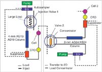
Figure 1: The experimental setup for matrix-elimination ion chromatography (MEIC).
An IonPac AS19 analytical column (4 × 250 mm) and an AG19 guard column (4 × 50 mm) were used for the first dimension analysis. The hydroxide eluent was generated from EGC KOH cartridge by feeding in a stream of deionized water. A continuously-regenerated anion trap column (CR-ATC) was used to remove any residual contaminants in the deionized water feed. An ASRS 300 4 mm suppressor was operated under external water mode. A PEEK tubing of 1000 μL was used as the sample loop. A carbonate removal device (CRD200 4 mm) was placed after the suppressor for removal of carbon dioxide from the carbonate peak in the sample. The addition of the CRD also helped to lower the amount of carbonate in the second dimension. The cell effluent was directed to the injection valve in the second dimension.
An IonPac AS24 analytical column (2 × 250 mm) and AG24 guard column (2 × 50 mm) were used for the second dimension analysis. The hydroxide eluent was generated from EGC KOH cartridge by feeding in a stream of deionized water. A CR-ATC was used to remove any residual contaminants in the deionized water feed. An ASRS 300 2 mm suppressor was operated under external water mode. A Carbonate Removal Device (CRD200 2 mm) was placed after the suppressor for removal of carbon dioxide from the carbonate peak in the sample. A UTAC-LP1 concentrator column was placed in the second injection valve for concentrating the target sample.
Reagents and Samples: Deionized water (18.2 MΩ cm, Millipore, Billerica, Massachusetts, USA) was used for eluent and sample preparation. Sodium bromate was purchased from Sigma-Aldrich (St Louis, Missouri, USA). A certified 1000 mg/L bromate solution was obtained from Ultra Scientific (N. Kingstown, Rhode Island, USA) and used as a secondary bromate standard.
Standard Solutions: A stock solution of 1000 mg/L bromate was prepared by dissolving 0.118 g of sodium bromate in DI water and diluting to 100 mL. The calibration standard solutions were prepared by serial dilution of the stock solution.
Ethylenediamine (EDA) is used as a preservative reagent for bromate and is added to the drinking water samples to remove any hypobromous acid in the drinking waters. A stock solution of 100 mg/L ethylenediamine was prepared by diluting 2.8 mL of EDA (Sigma-Aldrich) in DI water to a final volume of 100 mL. When the EDA stock solution is stored at 4 °C, the stock solution can be used for more than a month. Each sample and standard was spiked with 50 mg/L EDA by adding 50 μL of EDA stock solution into 100 mL of sample and standard solution.
High Ionic Water Matrix: The level of the matrix ions were chosen to represent a higher limit for most of the drinking water in the public drinking water systems. A synthetic high ionic water matrix, which consists of 100 mg/L chloride, 100 mg/L bicarbonate, 100 mg/L sulphate and 10 mg/L each of N (as nitrate) and P (as phosphate), was prepared as specified in the US EPA method 300.1 from the corresponding salts.
The drinking water samples were obtained from various public drinking water sources, following the standard EPA procedure, and stored at 4 °C. The bottled water sample was purchased from the grocery store and used without any treatment.
Results and Discussion
First Dimension Analysis of Bromate: The ideal first dimension column should provide good separation between the analyte and the matrix ions. For bromate analysis, chloride is the main peak eluting next to the bromate peak. If the two peaks elute too close to each other, there will not be an adequate window for heart-cutting the peak of interest. The presence of other ions will increase the overall ionic strength of the sample, leading to bromate retention time shift and peak broadening. The IonPac AS19 column (4 × 250 mm) was chosen as the first dimension column because it is a hydroxide-selective phase and a high capacity anion exchange column that was capable of handling the high matrix concentration. It provided good separation of bromate from chloride.
The benefit of choosing hydroxide as the eluent is because the suppressed effluent is water and it provides the best environment for concentrating and capturing the peak of interest into the concentrator column for further analysis in the second dimension. Additionally, the water background provides the best sensitivity for ion analysis.
Figure 2 shows the analysis of bromate with the IonPac AS19 column in the presence of various concentrations of matrix ions in a standard ion chromatography setup. As the matrix concentration increases, the retention time for the bromate peak shifts to earlier retention time, the peak also became broad along with a concomitant reduction in the peak height. These effects made the peak area integration a difficult task and affected the overall bromate quantification.
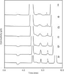
Figure 2: The effect of matrix concentration on the bromate peak shape. The matrix consists of chloride and sulphate and the concentration is: a) 0; b) 50 mg/L; c) 100 mg/L; d) 150 mg/L; e) 200 mg/L and f) 250 mg/L.
Cut Window Setting: For the MEIC approach, the cut window is defined here as the time window required for setting the valve timing to capture the peak of interest entirety into the concentrator column. The start of the cut window was determined by the injection of high ionic water matrix spiked with 15 μg/L bromate. Under this condition the bromate peak became a broad hump, and the front of the peak indicated the bromate peak started to elute off the first dimension column. Typically, the start of the cut window was set 0.4 min before the rise of the bromate peak. The end of the bromate peak was determined by the injection of 15 μg/L of bromate prepared in deionized water (termed as the reagent water sample). An additional 0.4 min was added to the end of the bromate peak to compensate for the system delay volume between the detector cell and the second injection valve. The overall cut window was 1.8 min wide for this setup. This cut window was adequate for the complete capture of the bromate peak from the suppressed effluent in the first dimension. Figure 3 shows an example of the cut window determination using the above method.
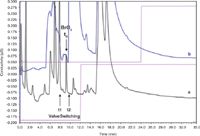
Figure 3: The first dimension analysis of the bromate peak in a) reagent water and b) high ionic water. The bromate concentration is 15 μg/L. The bromate retention time (tR) is 9.2 minutes. The start of the cut window (t1) is 8.0 minutes and the end of the cut window (t2) is 9.8 min.
The cut window should be monitored continuously with the first dimension analysis during the life of the column and adjusted accordingly to ensure complete capture of the bromate peak.
Second Dimension Analysis of Bromate: The ideal second dimension column should provide good separation between the bromate peak and any other contaminants. Since the majority of the matrix ions were already eliminated in the first dimension, the second column only needed to provide good separation between the bromate peak and other peaks that elute close to the bromate peak in the first dimension. The IonPac AS24 column was chosen for the second dimension analysis. This column is also a hydroxide-selective, high capacity anion exchange column. The column provided excellent separation between bromate and other peaks. Figure 4 shows the second dimension analysis of the bromate peak, both in reagent water and in the presence of high ionic water matrix. Most of the chloride peak was removed by the first dimension column, and the remaining small amount of chloride did not affect the bromate peak recovery and integration. This figure illustrates that the method provides consistent recovery even in the presence of matrix ions.
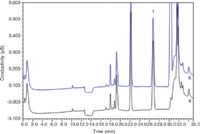
Figure 4: The second dimensional analysis of 15 μg/L bromate (Peak 1) in a) reagent water and b) high ionic water matrix. The bromate peak shows excellent recovery in both sample matrixes.
Table 1 shows the precision and accuracy of the present method for the two samples with a spike level of 0.5 μg/L and 5 μg/L of bromate respectively. The recovery values are well within the US EPA set limit of ±25% for samples containing bromate concentration within 5 times the minimum reporting level (MRL) and ±15% for those above five times the MRL.4

Table 1: Precision and accuracy.
The peak area obtained from the first dimension was also compared with the peak area from the second dimension. Table 2 listed the bromate peak area from the first and the second dimension analysis in reagent water. A larger peak area was obtained from the second dimension. The signal enhancement was approximately proportional to the ratio of the cross section area of these two column formats, yielding nearly a four-fold increase in sensitivity. Furthermore, this method is not limited to the 4 mm/2 mm column format shown here, and the second dimension can be lowered further to a 1 mm column format, or even a 0.4 mm capillary column format, thus is theoretically capable of providing further enhancement of the sensitivity.

Table 2: Response factor for 15 μg/L bromate between the first and second dimension analysis.
Method Performance: The method performance was evaluated by pursuing method linearity, MDL and precision and recovery tests.
The peak area response versus concentration was plotted for bromate in the concentration range of 0.15–15 μg/L in reagent water. A quadratic fit was used to fit the data as shown in Figure 5. A good correlation coefficient of 0.9997 was obtained. The method also showed very good method detection limit (MDL), precision and recovery. The MDL was determined by performing seven replicate injections of 0.2 μg/L bromate standard in reagent water. Using the calculation listed in US EPA method 300.1, the MDL for bromate is determined to be 0.037 μg/L. This is substantially lower than the detection limits reported in US EPA method 317.0 (0.12 μg/L) and 326.0 (0.17 μg/L), where a post-column derivatization approach was used to enhance the bromate detection.
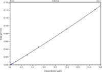
Figure 5: The calibration curve for bromate in the second dimension. The peak area response is plotted against the concentration using a quadratic fit.
Table 3 listed the precision and recovery data for a variety of real world drinking water samples and a bottle water sample. The samples were spiked with 0.5 μg/L and 5 μg/L bromate. The bromate recovery and method reproducibility was excellent as is shown from these results.
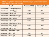
Table 3: The precision and accuracy of bromate analysis in various water samples spiked with 0.5 and 5.0 μg/L bromate.
The effect of the preservative agent ethylenediamine (EDA) was also investigated for the reagent water and high ionic strength matrix. Table 4 showed the comparison data between the sample spiked with EDA and without any EDA. Both samples showed good recovery with minimal impact on retention time. The stock EDA solution was maintained at 4 °C for a month and no noticeable effect was observed for the analysis.

Table 4: Effect of EDA on bromate retention time and recovery.
Overall the MEIC method was fully automated and analysis of a variety of samples with various matrix levels was feasible by the present method. The above method is presently used in US EPA method 302.0 and 314.2.20,22
Conclusion
In this article, we discussed a new application of the two dimensional MEIC method for bromate analysis. The method was able to eliminate a large amount of matrix ions and improve the detection of bromate in the presence of interfering matrix ions. The method is fully automated and showed excellent detection limit, precision and recovery. We have also demonstrated the use of the method for analysing various water samples and have demonstrated good recovery. The method can also be applied to other applications, where analysis of trace level analyte in the presence of large amount of matrix ions is desired.
Acknowledgements
We gratefully acknowledge the help and encouragement of H.P. Wagner from Lakeshore Engineering Services, Inc., B.V. Pepich from Shaw Environmental Inc., R. Joyce and B. De Borba from Dionex Corporation and D.J. Munch from US EPA throughout this study.
Rong Lin is a senior scientist at Dionex Corporation. He joined the company in 2000 and has been focusing on developing. new suppressor technologies and IC methods. He obtained his PhD in chemistry from University of California, Irvine in 2000.
Kannan Srinivasan has a PhD in Analytical Chemistry from the American University, Washington DC, USA. He was a guest scientist in the biotechnology division at National Institute of Standards and Technology, Gaithersburg, Maryland, USA from 1990 till 1993 when he joined the Dionex Corporation. He is currently a technical director in the R&D department. and is responsible for the development of novel consumable products for ion chromatography.
Christopher Pohl is senior vice president of R&D and chief science officer at Dionex Corporation. Christopher joined Dionex in 1979 where the focus of his work has been new stationary phase design. He received his BS in Analytical Chemistry from the University of Washington in 1973.
Reference
1. J.D. Pfaff, US EPA Method 300.0, Rev 2.1 (1993).
2. D.P. Hautman and D.J. Munch, US EPA Method 300.1, Rev 1.0 (1997).
3. Y. Liu et al., J. Biochem. Biophys. Methods, 60(3), 205–232 (2004).
4. H.P. Wagner et al., US EPA Method 317.0, Rev 2.0 (2001).
5. H.P. Wagner et al., US EPA Method 326.0, Rev 1.0 (2002).
6. H.P. Wagner et al., J. Chromatogr. A, 1039(1–2), 97–104 (2004).
7. R. Lin et al., Pittcon 2010, Technical Programme, 1960–1965.
8. S.R. Villasenor, J. Chromatogr. A, 602(1–2), 155–161 (1992).
9. C. Umile and J.F.K. Huber, J. Chromatogr. A, 723(1), 11–17 (1996).
10. S. Colombini, S. Polesello and S. Valsecchi, J. Chromatogr. A, 822(1), 162–166 (1998).
11. P.B.M. Caselli et al., J. Chromatogr. A, 1003(1–2), 133–141 (2003).
12. K. Tian, P.K. Dasgupta and T.A. Anderson, Anal. Chem., 75(3), 701–706 (2003).
13. K. Tian et al., Talanta, 65(3), 750–755 (2005).
14. S. Peldszus, P.M. Huck and S.A. Andrews, J. Chromatogr. A, 793(1), 198–203 (1998).
15. Y. Huang, S. Mou and Y. Yan, J. Liq. Chrom. & Rel. Technol., 22(14), 2235–2245 (1999).
16. Y. Huang, S. Mou and J.M. Riviello, J. Chromatogr. A, 868(2), 209–216 (2000).
17. K. Srinivasan, R. Lin and C. Pohl, IICS 2008 Scientific Programme, P32 (2008).
18. R. Lin et al., Analytica Chimica Acta, 567(1), 135–142 (2006).
19. H.P. Wagner et al., J. Chromatogr. A, 1155(1), 15–21 (2007).
20. H.P. Wagner et al., US EPA Method 314.2, Rev 1.0 (2008).
21. B. De Borba et al., J. Chromatogr. A, 1085(1), 23–32 (2005).
22. H.P. Wagner et al., US EPA Method 302.0, Rev 1.0 (2009).

Characterizing Plant Polysaccharides Using Size-Exclusion Chromatography
April 4th 2025With green chemistry becoming more standardized, Leena Pitkänen of Aalto University analyzed how useful size-exclusion chromatography (SEC) and asymmetric flow field-flow fractionation (AF4) could be in characterizing plant polysaccharides.


