Advances in Glycomics in Biology and Medicine
The importance of glycosylated structures in modern biology and medicine has been beyond dispute for many years, but there are still gaps in biochemical understanding. The current realization that virtually all major human diseases have been associated with glycosylation changes demands in-depth structural studies of these highly complex glycobiomolecules. Glycoscience with its many directions and a broad scope in both prokaryotic and eukaryotic systems is currently securing its place at the centre stage of modern biological research.
The importance of glycosylated structures in modern biology and medicine has been beyond dispute for many years, but there are still gaps in biochemical understanding. The current realization that virtually all major human diseases have been associated with glycosylation changes demands in-depth structural studies of these highly complex glycobiomolecules. Glycoscience with its many directions and a broad scope in both prokaryotic and eukaryotic systems is currently securing its place at the centre stage of modern biological research (1).
However, the enormous complexity of glycoconjugate molecules and the abundance of glycosylated proteins in biological fluids and tissues present significant challenges to modern analytical methods and measurement technologies. As underscored in the 2012 report of the National Research Council to the U.S. National Academies (2), developing new tools for glycoscience is one of the highest priorities of the general scientific inquiry.
The rapidly growing fields of glycomics and glycoproteomics reflect these on-going efforts. Biomolecular mass spectrometry (MS) is today the central identification and measurement technique for glycans and glycopeptides. The contemporary MS features state-of-theâart instrumentation in terms of ionization and fragmentation techniques, highly resolving mass analyzers, and very sensitive detection. However, the field of analytical glycobiology also urgently needs high-performance separation methods to assist MS measurements because of (a) the sheer complexity of the mixtures generated during various cleavages of glycoprotein molecules; and (b) glycan isomerism, which is both biologically relevant and methodologically difficult.
The role of separation scientists to develop better analytical methods and reliable protocols to study differences in glycosylation (for example, “normal” versus “aberrant” glycosylation levels) seems secure for a long time to come. Starting with sample preparation, fractionation, and preconcentration, and ending with the discrete resolution of glycoconjugates before MS measurements, many of these tasks are accomplished through chromatographic principles. Rapid advances are increasingly seen in many of these vitally important tasks of glycomics and glycoproteomics. Carbohydrate derivatization at microscale, such as permethylation or fluorescent labeling, are often desirable to enhance identification and measurements. Recent advanced methods in analytical glycoscience have been reviewed (3,4).
Glycomic profiling measurements have been particularly important in a search for disease biomarker candidates. Additionally, glycosylation analysis of therapeutic glycoproteins is becoming increasingly essential to evaluate their bioactivity, safety, immune response, and solubility. While deglycosylation protocols for N-linked glycans have been reliably developed during recent years, the same still cannot be said about quantitative analysis of O-linked oligosaccharides, although some progress has recently been made. The general profiling procedures may involve matrixâassisted laser desorption–ionization mass spectrometry (MALDI-MS), HPLC–fluorescence detection, capillary electrophoresis with laser-induced fluorescence (CE–LIF) detection, or capillary liquid chromatography tandem mass spectrometry (LC–MS/MS). Some of the MS techniques and CE–LIF can be complementary in yielding the information on glycan isomerism (5,6).
Our laboratory has had a long-term interest in the search for cancer biomarkers through glycomic comparative measurements (6,7) in the blood serum of patients with different disease conditions. More recently, we have extended our studies to glycomic profiling and structural characterization of urinary exosomes (8). Our current emphasis has also been on the isomerism concerning fucosyl substitution and sialyl linkages, in both N- and O-glycans, in relation to oncological conditions. With regards to N-glycans, typical profiles reflect high-mannose and complex-type oligosaccharides resulting from the major serum glycoproteins. It is now often the case that trace multiantennary N-glycans represent the very structures with important diseaseârelated information. To enhance their detection by MS techniques, we can use preconcentration based on the hydrophobicity of permethylated structures or the ion-exchange principles for multiply-sialylated structures. Certain amidation reactions can also be helpful in distinguishing sialyl linkage isomers in both capillary LC–MS/MS and microchip CE (6).
Exosomes originating from different tissues and encountered in biological fluids have recently received considerable attention for their diagnostic and therapeutic potential. Exosomes are extracellular nanoâsized vesicles encapsulating nucleic acids and proteins, both with and without glycosylation. There is increasing evidence that certain exosomes are involved in the cancer process, suppression of the immune system, drug resistance, and possibly metastasis. As a prelude to studying the exosome glycoconjugate composition in the genitourinary tract cancers, we have broadly characterized glycans (8) and proteins associated with urinary exosomes. The analytical profiles of N-glycans feature paucimannosidic structures, highâmannose, and large complexâtype structures. Using long capillaries packed with graphitized carbon black together with electrospray ionization MS/MS, we succeeded in partial resolution of some sialylated isomers of tetra-antennary glycans. This represents one of the most difficult analytical challenges in glycoscience (1).
References
- R.D. Cummings and J.M. Pierce, Chem.Biol.21, 1–15 (2014).
- National Research Council of the National Academies, Transforming Glycoscience: A Roadmap for the Future
- (The National Academies Press, Washington, D.C., USA, 2012).
- W.R. Alley Jr., B.F. Mann, and M.V. Novotny, Chem. Rev.113, 2668–2732 (2013).
- S. Gaunitz, G. Nagy, N.L.B. Pohl, and M.V. Novotny, Anal. Chem.89, 389−413 (2017).
- S. Mittermayr, J. Bones, and A. Guttman, Anal. Chem.85, 4228–4238 (2013).
- C.M. Snyder, W.R. Alley Jr., M.I. Campos, M. Svoboda, J.A. Goetz, J.A. Vasseur, S.C. Jacobson, and M.V. Novotny, Anal. Chem. 88(19), 9597–9605 (2016).
- W.R. Alley Jr., J.A. Vasseur, J.A. Goetz, M. Svoboda, B.F. Mann, D.E. Matei, N. Menning. A. Hussein, Y. Mechref, and M.V. Novotny, J. Proteome Res. 11(4), 2282–2300 (2012).
- G. Zou, J.D. Benktander, J.T. Gizaw, S. Gaunitz, and M.V. Novotny, Anal. Chem. 2017. DOI: 10.1021/acs.analchem.7b000629

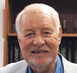
Milos V. Novotny, Department of Chemistry, Indiana University, Bloomington, Indiana, USA and Regional Centre for Applied Molecular Oncology Masaryk Memorial Oncological Institute, Brno, Czech Republic.

Silvia Radenkovic on Building Connections in the Scientific Community
April 11th 2025In the second part of our conversation with Silvia Radenkovic, she shares insights into her involvement in scientific organizations and offers advice for young scientists looking to engage more in scientific organizations.
Regulatory Deadlines and Supply Chain Challenges Take Center Stage in Nitrosamine Discussion
April 10th 2025During an LCGC International peer exchange, Aloka Srinivasan, Mayank Bhanti, and Amber Burch discussed the regulatory deadlines and supply chain challenges that come with nitrosamine analysis.
Removing Double-Stranded RNA Impurities Using Chromatography
April 8th 2025Researchers from Agency for Science, Technology and Research in Singapore recently published a review article exploring how chromatography can be used to remove double-stranded RNA impurities during mRNA therapeutics production.


