LC Europe: Overcoming Complex Analytical Challenges Posed by Targeted Multispecific Antibody Cancer Therapeutics
Complex multispecific antibodies overcome the limitations of standard monoclonal antibodies (mAbs), and have complex three-dimensional structures, greater heterogeneity, and often novel disulfide linkages and unusual post-translational modifications. As a result, the comprehensive characterization essential to the development of cost-effective manufacturing processes and safe and efficacious cancer treatments is much more challenging using traditional analytical instruments and techniques. Fortunately, there are new solutions available that provide fast results, often with greater sensitivity and resolution, even for these novel, complex molecules. The application of various new methods and instruments for the analysis of multispecifics is discussed as a means of highlighting their ability to facilitate the development of these important new modalities.
Targeted anti-cancer agents and immunotherapies have revolutionized the way in which cancer is treated today, but standard monoclonal antibodies (mAbs) do have limitations in selectivity and efficacy. Scientists are overcoming some of these issues through antibody engineering of whole mAbs or parts of mAbs fused to one another or other protein scaffolds. Incorporation of multiple binding sites or using specific fragments from conventional antibodies affords greater in vivo stability, more effective targeting, and greater efficacy.
Antibody Immunoglobulin | Image Credit: © freshidea - stock.adobe.com
Multispecific antibodies have complex three-dimensional structures and greater heterogeneity than traditional mAbs. They often include novel disulfide linkages and unusual post-translational modifications (PTMs). Consequently, the comprehensive characterization essential to the development of cost-effective manufacturing processes and safe and efficacious cancer treatments is much more challenging using traditional analytical instruments and techniques.
Full product quality assessment of multispecifics includes molecule identification, the identification of high-molecular-weight fragments and the percentage of heterodimers, determination of purity, charge heterogeneity and sequence variants, analysis of glycosylation and glycation, confirmation of molecular integrity, including sequence and disulfide linkage determination, and the identification of post-translational modifications, such as oxidation, deamidation, and isomerization.
New solutions are coming to market to provide faster results, greater sensitivity, and higher resolution, even for these novel, complex molecules. Fast glycan analysis using capillary electrophoresis (CE) with laser-induced fluorescence (LIF) detection facilitates characterization, quantitation, and sequencing of many glycans in peptibodies/fused proteins (1). Parallel processing of multiple samples simultaneously using multi-capillary CE approaches accelerates analysis and dramatically shortens new therapy development timelines, while still providing sensitive, high-resolution data (2). Rapid capillary zone electrophoresis (CZE) enables ultrafast charge variant analysis of next-generation antibodies with high-resolution (3).
Differential mobility separation (DMS) technology coupled with MS analysis eliminates the need for chromatography, thereby increasing the throughput of subunit analyses (4). New trapping and fragmentation technologies, meanwhile, enable MS analyses with increased sampling efficiency for greater fragment ion coverage and better sensitivity (2,5–7).
Materials and Methods
Fast Glycan Analysis with CE-LIF
For glycan analysis of the asparagine-linked oligosaccharides of the Fc-Fusion protein etanercept, carbohydrate release and APTS labeling was accomplished using the Fast Glycan Sample Preparation and Analysis kit (SCIEX) (1). CE-LIF was performed using the PA 800 Plus system (SCIEX), equipped with a solid-state laser-based fluorescent detector (λex=488 nm/λem=520 nm). A 20 cm effective length (30 cm total length; 50 μm I.D.) bare fused silica capillary was filled with the high resolution separation gel buffer (HR-NCHO) system. Pre-injection of water (3.0 sec/5.0 psi) was followed by 2.0 kV/2.0 sec sample injection and finally the introduction of the bracketing standard (2.0 kV/2.0 sec). The 32 Karat software, version 10.1 (SCIEX) was employed for data acquisition and analysis.
CE-Based Parallel Processing
Emicizumab (Genentech, Inc.) was analyzed on the BioPhase 8800 system from SCIEX with cartridges using the BioPhase CE-SDS Protein Analysis and cIEF kits (2). Data were acquired and processed using the BioPhase software, version 1.0.
Samples for CE-sodium dodecyl sulfate (SDS) were prepared following the standard SDS-molecular weight (MW) analysis assay kit protocol and analyzed (following capillary conditioning) at 25 °C using the BioPhase bare fused silica (BFS) capillary cartridge and UV absorbance detection at 220 nm.
For cIEF, emicizumab at a 5 mg/mL concentration was mixed with wide-range (W) and narrow-range (N) ampholytes at ratios of 7:1, 5:1, and 3:1 W/N (v/v) to improve the separation resolution of charge variants. Each sample mixture was mixed thoroughly at room temperature,and 100 μL aliquots were placed in the injecting well positions of the injection sample plate. Analysis was performed (after capillary conditioning) at 20 °C using the BioPhase neutral capillary cartridge and ultraviolet (UV) absorbance detection at 280 nm.
Ultrafast Charge-Variant Analysis with CZE
Ultrafast charge variant analysis of emicizumab (Genentech) and blinatumomab (Amgen) was performed on the PA 800 Plus system using the EZ-CE Pre-Assembled Capillary Cartridge and the CZE Rapid Charge Variant Analysis Kit (3). All data were acquired and processed by the 32 Karat software, version 10.1.
Emicizumab CZE charge heterogeneity analysis conditions were: 30 cm total capillary length (20 cm effective, 50 μm ID); applied electric field strength: 1000 V/cm; separation temperature: 25 °C; and pressure injection at 5 psi for 10 sec. The blinatumomab sample (0.2 mg/mL) was washed three times with 100 μL of water through a 10 kDa cut-off centrifugation filter (Millipore) at 14 000 x g to eliminate matrix-related effects. Separation conditions were the same, with the exception of pressure injection at 5 psi for 20 sec.
Three-day forced degradation of emicizumab (1 mg/mL protein solution in 100 mM Tris-HCl buffer) was performed at pH 8.7 and 45 ºC.
MS Using Differential Mobility Separation (DMS) Technology
A sample (10 mg/ml) of a bsAb (Merck Healthcare KGaA) was reduced at room temperature using tris(2- carboxyethyl)phosphine then diluted to a final concentration of 0.1 mg/mL in 10% acetonitrile/1% formic acid (v:v) (4). Chromatography (10 μL/1 μg injection) was performed on a MassPREP desalting column (2.1 mm × 5 mm, 20 μm, 1000 Å) at 80 °C using two mobile phases (A: Water + 0.1% formic acid, B: acetonitrile + 0.1% formic acid) with a flow rate of 500 μL/min. MS was performed using an ExionLC system coupled to a TripleTOF 6600 system equipped with an IonDrive Turbo V source and SelexION DMS device (temperature: 150 °C; separation voltage: 3500 V; compensation voltage: -9 / -3 / 9 V). Data were processed using SCIEX OS software, version 1.6.1 with the BioToolKit and BPV Flex 2.0 software.
MS Using Zeno Trapping and Tunable Electron Fragmentation Technologies
For peptide mapping of a trispecific antibody (5), the sample was denaturated with 7.2 M guanidine hydrochloride in 100 mM Tris buffer pH 7.2, followed by reduction with 10 mM DL-dithiothreitol and alkylation with 30 mM iodoacetamide. Digestion was performed with trypsin/Lys-C enzyme at 37 °C for 16 h. Chromatography was performed using a CSH C18 column Waters ACQUITY CSH C18 column (2.1 × 100 mm, 1.7 μm, 130 Å) at 50 ºC using an ExionLC system, with 10 μL (4 μg) of the trypsin/Lys-C digest separated. Mobile phases A and B consisted of 0.1% formic acid in water and acetonitrile, respectively. A 60-min gradient profile was used at a flow rate of 300 μL/min. MS data were acquired with a data-dependent acquisition (DDA) method using the ZenoTOF 7600 system with either CID or EAD as fragmentation mode and processed with Byos software (Protein Metrics Inc.). General method parameters were kept the same between the two methods, but the electron energies for the CID and EAD cells were rolling and 7 eV, respectively.
Peptide mapping of etanercept (reference material #8671, NIST) (6) was performed on a sample prepared in the same manner, except that the digest was further treated with an endoglycosidase (PNGase F, Agilent Technologies Inc.) for removal of all N-glycans followed by reaction with SialEXO (Genovis Inc.) to cleave sialic acids. For chromatography, the same column, instrument, and mobile phases were used, but the sample size was 3 µL (4 µg), the column temperature was 60 ºC and the flow rate was 250 μL/min. MS data were acquired using a 10 Hz DDA method on the ZenoTOF 7600 system with the electron energy for the EAD cell set to 7 eV. The data were processed with Byos software (Protein Metrics Inc.).
Results and Discussion
Glycan Characterization, Quantitation, and Sequencing
Accurate glycan analysis is essential because glycan microheterogeneity, charge heterogeneity, and product- related impurities impact the safety, stability, and efficacy of antibody-based therapeutics. Typical LC- and CE-based methods require labor intensive, time-consuming sample preparation including glycan release, purification (multiple centrifugation steps), fluorophore labeling, and pre-concentration prior to analysis.
A new method that uses a high-resolution gel (HR-NCHO) buffer enables excellent separation performance even with a simple preparation protocol that requires just one hour for glycan release, fluorophore labeling, and clean-up and 5 min for CE-LIF analysis of the fluorophore-labeled glycans (8). The protocol leverages a novel magnetic bead-mediated process that affords reproducibly high yields (9,10). Because there is no need for centrifugation or vacuum-centrifugation, the method is suitable for single analyses, and can also be used with automated liquid handling platforms that process up to 96 samples at a time (11). Use of a triple-internal standard approach and specialized software allows structural assignment of the separated glycans (12).
The utility of the method for assessment of N-glycan microheterogeneity was demonstrated for the National Institute of Standards and Technology monoclonal antibody (NISTmAb) reference material (RM 8671) (13).
Glycan analysis of the asparagine-linked oligosaccharides of the Fc-Fusion protein etanercept (APTS and 2-AB labeled etanercept N-glycans) using the fast glycan method was also compared to analysis using traditional ultrahigh-pressure LC (UHPLC) with fluorescence (FL) detection (1). Etanercept has three N-linked glycosylation sites, one at the Fc domain and two at the TNFα receptor domain of the molecule. The UHPLC analysis took nearly 40 min and identified 12 glycan structures. The CE-LIF analysis revealed 18 glycans with fast separation in <3.5 min. Even better resolution could be achieved for the CE-LIF analysis when a longer capillary (50 cm effective length) was employed.
CE-Based Parallel Processing
CE offers high-resolution and robust separation of molecules and is an invaluable technique for the characterization of complex biotherapeutics. Sodium dodecyl sulfate capillary gel electrophoresis (SDS-CGE, also referred to as CE-SDS) can monitor protein size variants caused by artificial fragments resulting from insufficient alkylation (14). With temperature optimization, it is possible to fine tune the separation performance for even complex multispecifics (15). cIEF can monitor protein charge variants caused by PTMs that are considered CQAs due to their influence on protein stability and potency (16). Indeed, cIEF can separate charge variants with very close isoelectric point (pI) values and is applied in quality control (QC) and lot-release tests in the biopharmaceutical industry (17).
The ability to process multiple CE samples simultaneously while still benefitting from the high resolving power and excellent quantitation and reproducibility of CE can accelerate method and process development for multispecifics (2). Parallel processing allows the implementation of a design-of-experiment (DoE) approach for the development of an optimized CE-based method. It also enables more rapid analysis of larger quantities of samples generated during DoE studies pursed as part of process development activities. If the multiplexed CE system enables seamless transfer of optimized methods to single-analysis instruments employed for QA/QC and thus in a GMP environment, scaleup can also be accelerated.
To demonstrate the value of parallel CE processing, comprehensive characterization of emicizumab, a biospecific antibody (bsAb) used for the treatment of hemophilia A, was performed via CE-SDS and cIEF using a multi-capillary CE system (2). With the employed workflow, CE was leveraged to achieve standard characterization of protein size and charge variants using a one-platform system with high reproducibility and throughput.
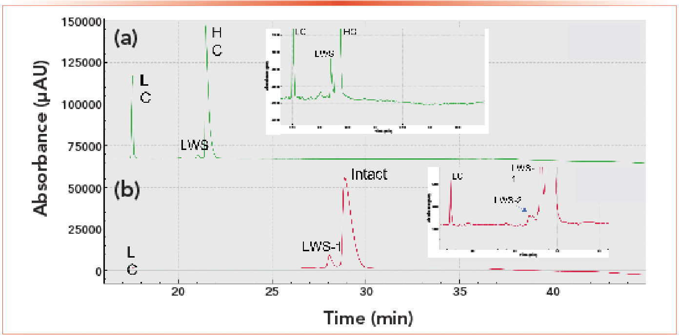
Emicizumab has an asymmetric structure with heterogeneous heavy chains (HC) and a common light chain (LC). Reduced and nonreduced forms were analyzed for purity using CE-SDS (Figure 1). The heterogenous HCs were not separated, as indicated by a single peak in the electropherogram, and no high-molecular-weight species (HWS) were detected. The electropherogram of the reduced form revealed a low-molecular-weight species (LWS) that could be the non-glycosylated heavy chain. The intact form emicizumab (HHLL) was determined to be greater than 92%, and the LC to HC was approximately 2:5.
Figure 1: CE-SDS separation of emicizumab. (a) Green shows reduced, while (b) red shows intact forms. Both are analyzed by CE-SDS.
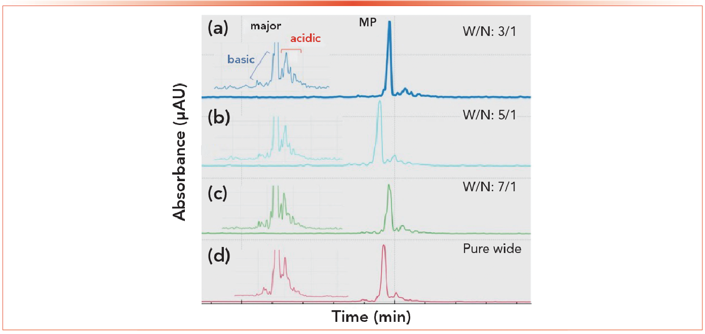
Excellent repeatability was observed for both SDS and cIEF analyses of emicizumab across capillaries and injection replicates, expediting the optimization of parameters that can affect separation performance. The pI values of the charge variants of emicizumab lies in the range of 6–7, indicating that a narrow range ampholyte of 6–8 can help improve separation resolution. Various combinations of wide-range ampholytes (3–10) (W) and narrow-range ampholytes (6–8) (N), including W/N of 3/1, 5/1, and 7/1 (v/v) were tested for resolution improvement. Figure 2 shows that N led to improved resolution but increased noise.
Figure 2: (a-d) Improving cIEF separation resolution with various combinations of ampholyte.
As a next step, full factorial DoE with 4 factors (pH and concentration of the anolyte, catholyte, and chemical mobilizer solutions and concentrations of urea and cathodic and anodic stabilizers), each with 2 input levels, would require 16 runs for optimization. On a single-capillary system that would take approximately 18 h to complete, whereas with a multiplexed system with eight capillaries, only two runs would be necessary.
Ultrafast Charge-Variant Analysis with CZE
Charge heterogeneity analysis of multispecifics is important during product development, production, stability, and release testing (18). As many as 15 variants can be generated in a single batch of a multispecific antibody, with perhaps two of them being therapeutically relevant. Any method for charge heterogeneity analysis must therefore be high resolution, rapid, accurate, and reproducible. Commonly used ion exchange chromatography and isoelectric focusing techniques tend to be relatively slow, however (19,20).
CZE using nanoliter injection volumes separates analytes based on their charges and hydrodynamic volume-to-charge ratios (3). It can be used for the separation of large and small molecules with a wide range of chemical and physical properties and offers plate numbers approximately several dozen times greater than that of high performance liquid chromatography (HPLC). Analysis times are typically 10 min for next-generation protein therapeutics in their native forms.
When the bsAb emicizumab was diluted with water to a concentration of 1 mg/mL and six consecutive CZE analysis runs were completed in less than 8 min each, six peaks were observed (two faster-migrating, possibly basic species, three slower-migrating, possibly acidic species, and the main peak for the bsAb) with high reproducibility (3).
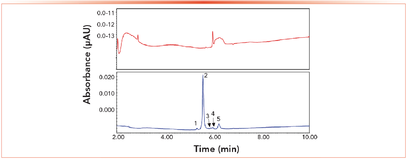
A low-concentration (0.2 mg/mL) sample of the BiTE bsAb blinatumomab required buffer exchange prior to injection for CZE analysis due to interference of the initial buffer (3). Five peaks were revealed for this bsAb (Figure 3). Since CZE runs typically take < 10 min, it is recommended that analysis first be performed without any sample preparation, and if insufficient separation is observed, a simple buffer exchange (washing with water) should eliminate any matrix effects.
Figure 3: (a) CZE charge heterogeneity analysis of the bsAbs emicizumab (with formulation buffer matrix); (b) CZE charge heterogeneity analysis of blinatumomab (buffer exchange using 10 kDa cutoff buffer).
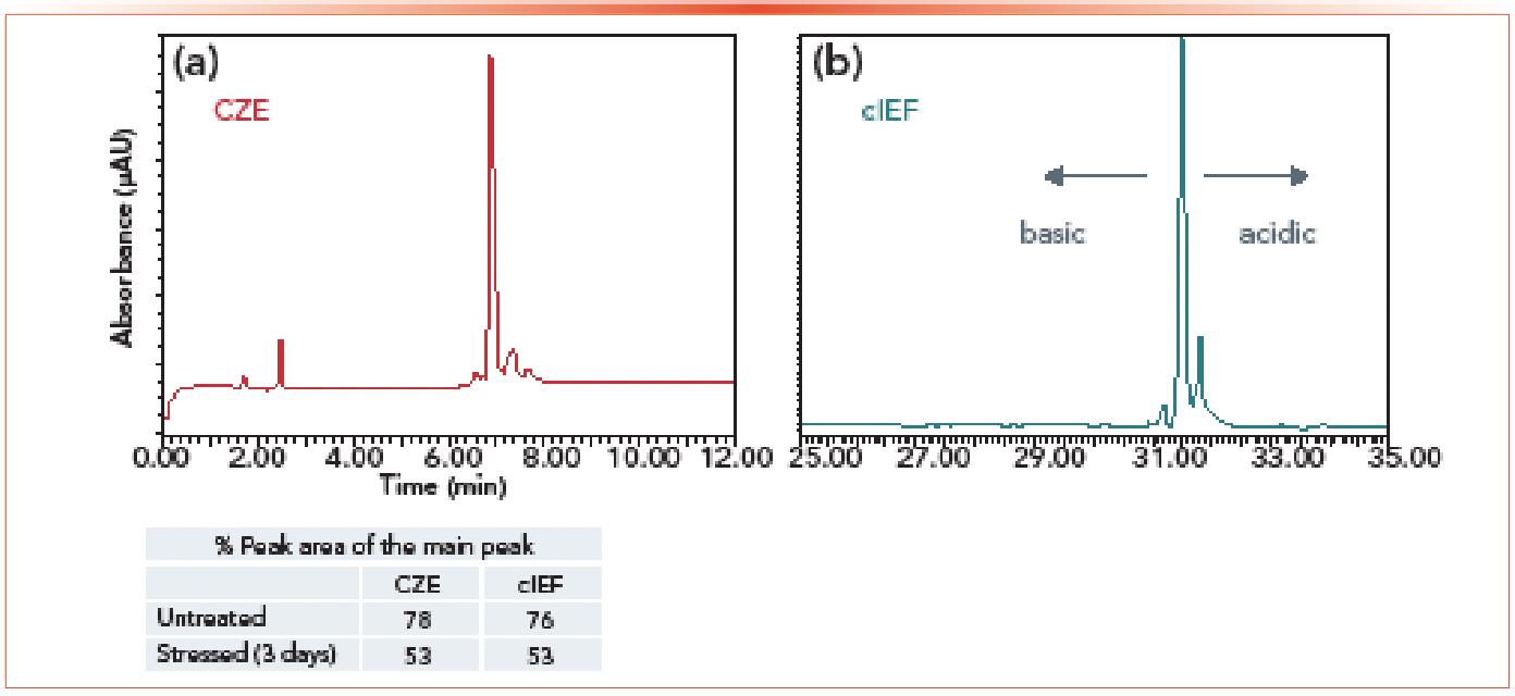
A three-day forced degradation study of emicizumab at pH 8.7 and 45 °C was also performed (3). CZE and cIEF analyses were both performed (Figure 4), because cIEF can be used to determine isoelectric point (PI) values (although the run time for cIEF is typically 30–40 min). These results demonstrated that CZE can be readily applied to rapid, high-throughput charge heterogeneity profiling; results were obtained five times faster than those for cIEF with apparently very similar relative peak areas for the main peak.
Figure 4: This figure shows (a) CZE vs. (b) cIEF quantification of the forced deamidation of emicizumab.
It should also be noted that, when combined with MS, the identification of deamidated peptides following separation from their non-deamidated counterparts can be achieved, which is not possible with LC (3).
MS-Based Subunit Analysis with Increased Throughput Using Differential Mobility Separation (DMS) Technology
Subunit analysis by LC–MS enables the analysis of the individual chains of bsAbs in a single method, but the chromatographic separation of protein chains prior to MS analysis is challenging and can complicate data analysis. One solution for overcoming this issue is to use differential mobility separation (DMS) technology coupled with MS analysis. This approach was demonstrated for a bispecific protein sample consisting of three distinct amino acid chains (one light chain, one AG chain, and one GA chain) forming two different binding domains (4).
Chromatographic separation of the chains required a run time greater than 20 min. The use of DMS following installation of the DMS cell, which required just 2 min, enabled unambiguous characterization of each chain in a single injection without chromatography for simpler data collection and analysis and reduced analysis time for increased throughput.
For this bsAb, the three different protein chains were separated by serially scanning three compensating voltages (CoVs) within a single injection (4). The total ion chromatograms for each CoV could be visualized separately and the raw data evaluated, providing clean spectra and giving more confidence in the interpretation of the reconstructed results.
New Zeno Trapping and Tunable Electron Fragmentation Technology Impacts
In traditional time-of-flight mass spectrometry (TOF-MS) instruments, the duty cycle problem arises because fragment ions arrive at the pulser region with a spread of velocities, and only a small percentage get accelerated into the flight tube. With Zeno trap, the ion fragments are trapped and then ejected in order and inversely proportional to their m/z values so that all of them arrive at the TOF pulser simultaneously. More than 90% are injected into the TOF, leading to improved MS/MS ion sampling for all m/z, and particularly for low m/z species (10X or more). The result is greater sensitivity for MS/MS and high-resolution multi-reaction monitoring (MRMHR) acquisition scan modes and greater ion fragment coverage (5).
Meanwhile, unlike traditional electron capture dissociation (ECD) for MS applications, which relies solely on electrons with low kinetic energy (0 EV), controlled electron activated dissociation (EAD) enables tuning of the velocity of the electrons, and thus their kinetic energy (5). Not only is electron transfer dissociation at 0 EV possible, but also “hot” ECD mechanisms at 7 EV for peptide mapping of glycopeptides and disulfide-bonded peptides with side-chain preservation for isomer confirmation, as well as electron impact excitation of ions (EIEIO) at 10 EV for analysis of small-molecule organics. Notably, EAD provides similar quantitative performance and sequence data compared to collision-induced dissociation (CID), but with improved fragment ion coverage (6). The EAD reaction is also robust, reproducible, and quantitative and suitable for use with liquid chromatography.
When Zeno trapping is combined with EAD fragmentation, much greater fragmentation coverage and increased sensitivity are realized at fast scan rates of up to 133 Hz, even for data-dependent and MRMHR acquisitions (5).
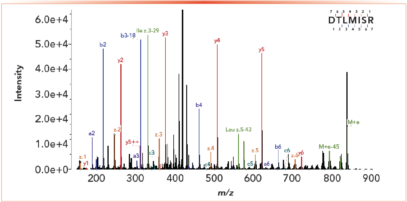
Peptide mapping of a trispecific candidate with a main binding site and two single-chain variable fragments (SCFVs) with affinity to secondary and tertiary targets shows the value of the combined use of Zeno trapping and EAD fragmentation (5). Confirmation of leucines and isoleucines, even though they differ by just 14 Da, was achieved (Figure 5), which otherwise would require the use of time-consuming and often inconclusive methods, such as DNA sequencing and Edman Degradation (5). It was also possible to resolve aspartic and isoaspartic acids (the result of deamidation of asparagine), which can impact target binding.
Figure 5: Figure showing the MS spectrum of a peptide from a trispecific antibody with both leucine and isoleucine fragments.
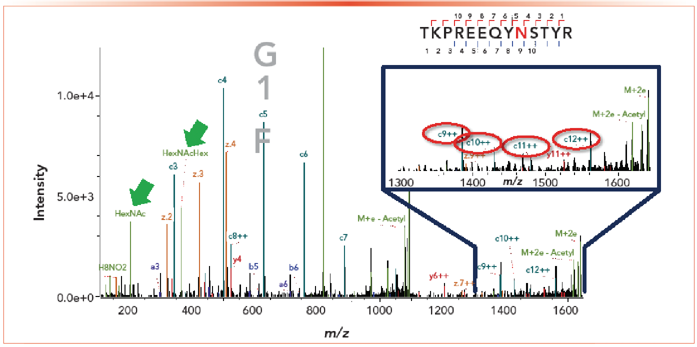
Localization data for labile molecules, such as glycans, can be difficult to obtain using CID fragmentation. With EAD, however, the integrity of labile modifications is maintained, allowing for generation of both localization and identity information. As can be seen in Figure 6, confirmation of the site of glycosylation was possible for the trispecific candidate. Detection of oxonium ions helped to confirm the identity of the glycopeptide (5).
Figure 6: Figures showing the MS spectrum of a peptide from the trispecific antibody with intact glycans. Confirmation of the site of glycosylation was possible for the trispecific candidate. Detection of oxonium ions helped to confirm the identity of the glycopeptide.
Analysis of disulfide bonds was also simplified. Nonreducing sulfide mapping of multispecifics generally results in large peptides that require additional fragmentation often not achievable with CID, but for which full sequence coverage is possible using EAD with the hot ECD mechanism (5). In the case of the trispecific candidate, 7,000 DA and 11,000 DA peptides were analyzed with excellent coverage across the entire peptides. In addition, peaks for the individual peptides that make up the larger disulfide-bonded peptides were observed.
EAD was also used to confirm the existence of disulfide and trisulfide linkages as well as free thiols in a bsAb in which two different heavy chains and two identical light chains are linked (6). An EAD platform method was developed that provides signature fragments to differentiate disulfide and trisulfide linkages, enabling a higher level of structural characterization in one injection. Increased detection of fragments (by 5- to 10-fold) enabled free thiol detection at low abundances. The reproducibility of EAD fragmentation also enabled analysis of more precursors than other alternative and low-reproducibility fragmentation techniques.
While both EAD and CID were able to identify all expected disulfide linkages from the bsAb, EAD provided higher MS/MS coverage for long peptides (6).
Conclusions
Multispecific antibodies are complex molecules with potential to serve as highly effective, targeted therapies. Ensuring their structural integrity, purity and safety is essential, but comprehensive characterization can be challenging given their greater structural complexity and heterogeneity. The time constraints often associated with accelerated development timelines for breakthrough address the developability gap by more rapidly identifying candidates with the greatest likelihood of success with respect to efficacy, safety, and manufacturability.
Analytical tools that can provide robust, reliable, in-depth information on critical quality attributes in a high-throughput manner are needed so that more detailed data can be gathered to enable informed decision-making at early development stages. Fast glycan analysis, parallel CE processing, CZE, MS leveraging DMS technology, and the use of Zeno trapping and EAD for MS analyses are all examples of advanced analytical techniques designed to enable rapid and robust analyses of even complex multispecifics.
References
(1) Guttman A.; Szigeti, M. Comparative Characterization of the Fc Domain N-glycosylation in Monoclonal Antibody and Fusion Protein Therapeutics by CGE-LIF and UPLC-FL. SCIEX Separations, Brea, CA; Horváth Csaba Laboratory of Bioseparation Sciences, University of Debrecen, Hungary, 2018. https://sciex.com/content/dam/SCIEX/pdf/tech-notes/all/CE_vs_UHPLC.pdf (accessed 2023-02-15).
(2) Yang, Z.; Santos, M.; Li, T.; Mollah, S. One-Platform Strategy for Comprehensive Characterization of the Bispecific Antibody, Emicizumab, Using the BioPhase 8800 System. SCIEX, Brea, CA, 2023. https://sciex.com/tech-notes/ce/One-platform-strategy-for-comprehensive-characterization-of-bispecific-antibody-emicizumab-using-multi-capillary-BioPhase-8800-system (accessed 2023-02-15).
(3) Guttman, A. CZE: Ultrafast Charge Variant Analysis for Next-Generation Antibody Therapeutics. SCIEX, Brea, CA, 2021. https://www.chromatographytoday.com/article/bioanalytical/40/sciex/cze-ultrafast-charge-variant-analysis-for-next-generation-antibody-therapeutics/2948 (accessed 2023-02-15).
(4) Pohl, K.; Schanz, J.; Tonillo, J.; Mehl D.; Kellner, R.; McCarthy, S. Increased Throughput for the Characterization of Bispec ific Antibodies on the Subunit Level Using SelexION Differential Mobility Separation (DMS) Technology. SCIEX, Framingham, MA, USA; Merck Healthcare KGaA, Darmstadt, Germany; SCIEX, Villebon sur Yvette, France, 2020. https://sciex.com/content/dam/SCIEX/pdf/tech-notes/all/selexion-bispecific-antibody.pdf (accessed 2023-02-15).
(5) Mahan, A. Accelerating the Characterization of Next-Generation Biotherapeutics with Mass Spectrometry Leveraging Novel Ion Trapping and Alternative Fragmentation Technologies. SCIEX, Brea, CA, 2021. https://www.chromatographyonline.com/view/acclerating-the-characterization-of-next-generation-biotherapeutics-with-novel-ion-trapping-and-alternative-fragmentation (accessed 2023-02-15).
(6) Zhang, Z. Comprehensive Analysis of Next Generation Biologics Using Advanced Data Interpretation Software. SCIEX, Brea, CA, 2021. https://www.labroots.com/webinar/biotherapeuric-characterization-biologics-explorer-software2 (accessed 2023-02-15).
(7) Bi, X. Comprehensive Characterization of Etanercept Using the ZenoTOF 7600 System. SCIEX, Brea, CA, 2021. https://www.labroots.com/webinar/comprehensive-characterization-etanercept-using-zenotof-7600-system (accessed 2023-02-15).
(8) Guttman, A.; Szigeti, M.; Lou, A.; Gutierrez, M. Fast Glycan Labeling and Analysis: High-Resolution Separation, Quantification, and Identification in Minutes. SCIEX, Brea, CA; Horváth Csaba Memorial Laboratory of Bioseparation Sciences, University of Debrecen, Hungary, 2017. https://sciex.com/content/dam/SCIEX/pdf/tech-notes/all/Fast-Glycan-Labeling-and-Analysis-High-Resolution-Separation-and-Identification-in-Minutes.pdf (accessed 2023-02-15).
(9) Váradi C.; Lew C.; Guttman A. Rapid Magnetic Bead Based Sample Preparation for Automated and High Throughput N-glycan Analysis of Therapeutic Antibodies. Anal. Chem. 2014, 86 (12), 5682–5687. DOI: 10.1021/ac501573g
(10) Szigeti M.; Lew C.; Roby K.; Guttman A. Fully Automated Sample Preparation for Ultrafast N-Glycosylation Analysis of Antibody Therapeutics. J. Lab Autom. 2016, 21 (2), 281–286. DOI: 10.1177/2211068215608767
(11) Candish, E.; Gutierrez, M.; Szigeti, M.; Guttman, A. Fast Glycan Sequencing Using a Fully Automate Carbohydrate Sequencer. SCIEX, Brea, CA; Horváth Csaba Memorial Laboratory of Bioseparation Sciences, University of Debrecen, Hungary, 2017. https://sciex.com/content/dam/SCIEX/pdf/tech-notes/all/single-analytical-platform-for-glycan-analysis-charge-heterogeneity.pdf (accessed 2023-02-15).
(12) Kovacs, Z.; Szarka, M.; Szigeti, M.; Guttman, A. Separation window dependent multiple injection (SWDMI) for large scale analysis of therapeutic antibody N-glycans. J. Pharm. Biomed. Analysis 2016, 128, 367–370. DOI: 10.1016/j.jpba.2016.06.002
(13) Jarvas G.; Szigeti M.; Chapman J.; Guttman A. Triple-Internal Standard Based Glycan Structural Assignment Method for Capillary Electrophoresis Analysis of Carbohydrates, Anal Chem. 2016, 88 (23), 11364–11367. DOI: 10.1021/acs.analchem.6b03596
(14) Candish, E.; Gutierrez, M.; Szigeti, M.; Guttman, A. Fast Glycan Sequencing Using a Fully Automate Carbohydrate Sequencer; SCIEX, Brea, CA; Horváth Csaba Memorial Laboratory of Bioseparation Sciences, University of Debrecen, Hungary, 2017. https://sciex.com/content/dam/SCIEX/pdf/tech-notes/all/single-analytical-platform-for-glycan-analysis-charge-heterogeneity.pdf (accessed 2023-02-15).
(15) Wagner E.; Colas O.; Chenu S.; Goyon A.; Murisier A.; Cianferani S.; François Y.; Fekete S.; Guillarme D.; D’Atri V.; Beck A. Determination of Size Variants by CE-SDS for Approved Therapeutic Antibodies: Key Implications of Subclasses and Light Chain Specificities. J. Pharm. Biomed .Anal. 2020, 184, 113166. DOI: 10.1016/j.jpba.2020.113166
(16) Guttma, A.; Filep, C. Better Separation Resolution of New Biopharmaceutical Modalities through Fine Tuning of the Temperature with CE-SDS; SCIEX, Brea, CA; Horváth Csaba Memorial Laboratory of Bioseparation Sciences, University of Debrecen, Hungary. https://sciex.com/tech-notes/ce/better-separation-resolution-of-new-biopharmaceutical-modalities (accessed 2023-02-15).
(17) Liu, H.; Ponniah, G.; Zhang, H.M.; Nowak, C.; Neill, A.; Gonzalez-Lopez, N.; Patel, R.; Cheng, G.; Kita, A.Z.; Andrien, B. In vitro and in vivo Modifications of Recombinant and Human IgG Antibodies. MAbs 2014, 6 (5), 1145–1154. DOI: 10.4161/mabs.29883
(18) Cao, M.; De Mel, N.; Shannon, A.; Prophet, M.; Wang, C.; Xu, W.; Niu, B.; Kim, J.; Albarghouthi, M.; Liu, D.; Meinke, E.; Lin, S.; Wang, X.; Wang, J. Charge Variants Characterization and Release Assay Development for Co-formulated Antibodies as a Combination Therapy. MAbs 2019, 11 (3), 489–499. DOI: 10.1080/19420862.2019.1578137
(19) Singh, S.K.; Narula, G.; Rathore, A.S. Should Charge Variants of Monoclonal Antibody Therapeutics be Cnsidered Critical Quality Attributes? Electrophoresis 2016, 37 (17–18), 2338–2346. DOI: 10.1002/elps.201600078
(20) Stoll, D.; Danforth, J.; Zhang, K.; Beck, A. Characterization of Therapeutic Antibodies and Related Products by Two-Dimensional Liquid Chromatography Coupled with UV Absorbance and Mass Spectrometric Detection. J. Chromatography 2016, 1032, 51–60. DOI: 10.1016/j.jchromb.2016.05.029
About the Author
Mark Lies is the Sr. Global Market Development Manager of Protein, mAbs, and ADC at SCIEX.
Extracting Estrogenic Hormones Using Rotating Disk and Modified Clays
April 14th 2025University of Caldas and University of Chile researchers extracted estrogenic hormones from wastewater samples using rotating disk sorption extraction. After extraction, the concentrated analytes were measured using liquid chromatography coupled with photodiode array detection (HPLC-PDA).
Silvia Radenkovic on Building Connections in the Scientific Community
April 11th 2025In the second part of our conversation with Silvia Radenkovic, she shares insights into her involvement in scientific organizations and offers advice for young scientists looking to engage more in scientific organizations.
Regulatory Deadlines and Supply Chain Challenges Take Center Stage in Nitrosamine Discussion
April 10th 2025During an LCGC International peer exchange, Aloka Srinivasan, Mayank Bhanti, and Amber Burch discussed the regulatory deadlines and supply chain challenges that come with nitrosamine analysis.


