Fundamental and Practical Aspects of Liquid Chromatography and Capillary Electromigration Techniques for the Analysis of Phenolic Compounds in Plants and Plant-Derived Food, Part 1: Liquid Chromatography
LCGC Europe
Column-based liquid phase separation techniques, such as liquid chromatography (LC) in reversed phase separation mode and capillary electromigration techniques, using continuous electrolyte systems, are widely used for the identification and quantification of phenolic compounds in plants and food matrices of plant origin. This paper is the first of a two-part review article discussing fundamental and practical aspects of both LC and capillary electromigration techniques used for the analysis of phenolic compounds occurring in plant-derived food and in edible and medicinal plants. The chemical structure and distribution of the major phenolic compounds occurring in the plant kingdom, as well as the main methods used for their extraction and sample preparation, are also discussed. Part 1 will focus on liquid chromatography.
Photo Credit: Skyler Ewing/Shutterstock.com

Column-based liquid phase separation techniques, such as liquid chromatography (LC) in reversed phase separation mode and capillary electromigration techniques, using continuous electrolyte systems, are widely used for the identification and quantification of phenolic compounds in plants and food matrices of plant origin. This paper is the first of a two-part review article discussing fundamental and practical aspects of both LC and capillary electromigration techniques used for the analysis of phenolic compounds occurring in plant-derived food and in edible and medicinal plants. The chemical structure and distribution of the major phenolic compounds occurring in the plant kingdom, as well as the main methods used for their extraction and sample preparation, are also discussed. Part 1 will focus on liquid chromatography.

Plant secondary metabolites, also known as specialized metabolites, are organic compounds produced along with the primary biosynthetic and metabolic routes that are believed to be mainly produced in response to the interactions of the plant with the environment.
These compounds protect the plant against biotic and abiotic stresses, including pathogens, predators, ultraviolet light, and drought (1). In addition, secondary metabolites may confer specific sensory characteristics to food products and play important roles in disease prevention and healthâpromoting effects of edible plants and plantâderived food products (2–5). Based on their biosynthetic origins, plant secondary metabolites are usually classified into three major classes: terpenoids, alkaloids, and phenolic compounds, the last of which are one of the most important and widespread class of secondary metabolites in the plant kingdom (6).
Phenolic compounds form an integral part of the human diet, contributing to the sensorial properties of food products and to the beneficial effects of the Mediterranean diet on human health, mainly as a result of their antioxidant properties (7). These compounds, as well as many other plant secondary metabolites, are also important as bioactive components in medicinal plants and have numerous biological activities and a variety of health benefits for chronic and degenerative human diseases (8).
Important steps for the assay of phenolic compounds in plants and plant-derived food are sample preparation and extraction, followed by identification and quantification using various instrumental analytical methods, most of which use column-based high performance liquid phase separation techniques coupled to a suitable detection method. Traditionally, total phenolic compounds are determined using spectrophotometric methods based on the Folin-Ciocalteau reaction (9), which generate a coloured product as the result of the oxidative titration of phenolate anions by phosphotungstate and phosphomolybdate. More sophisticated methods, based on instrumental analytical techniques, are needed to identify and quantify each of the main phenolic compounds present in a given sample. Among them, liquid chromatography (LC), generally in the reversed phase separation mode, is the technique of choice, while capillary electrophoresis (CE) is gaining increasing acceptance because of its high separation efficiency, short analysis time, and extremely small sample and reagent volume requirements.
This is the first of a two-part review article aimed at discussing fundamental and practical aspects of both LC and capillary electromigration techniques used for the analysis of phenolic compounds occurring in edible and medicinal plants and in plant-derived food and dietary supplements. Part 1 will focus on liquid chromatography of these compounds and the chemical structure and distribution of the major phenolic compounds, as well as the main methods used for sample preparation and extraction.
Major Phenolic Compounds
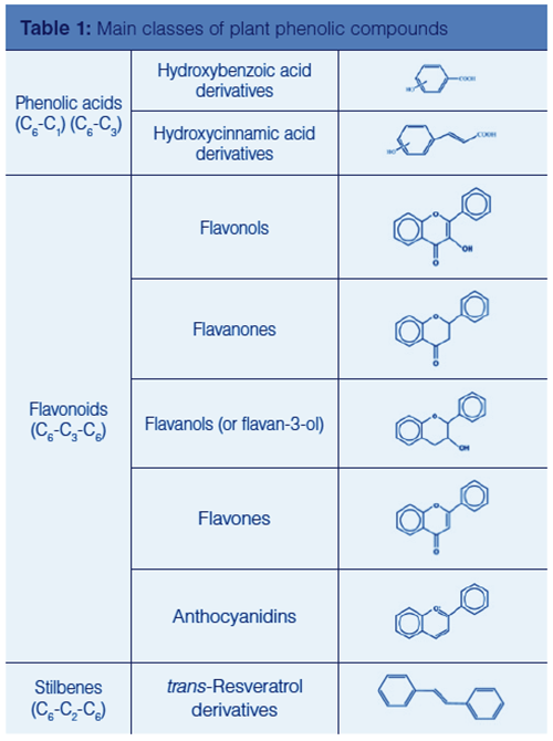
Phenolic compounds, also referred to as phenolics, comprise a large number of heterogeneous structures that range from simple molecules to highly polymerized compounds, which are commonly bound to other molecules, frequently to sugars, although phenolics in free form also occur. Among glycosylated phenolic compounds, both C- and O-glycosylations are found. A common structural characteristic of phenolics is the presence of at least one aromatic ring hydroxyl-substituted. They are commonly divided into different subclasses according to the number of aromatic rings, the structural elements that bind these rings to each other, and the substituents linked to the rings (see Table 1).

The simplest form of phenolics are the phenolic acids, which can be divided into benzoic acid and cinnamic acid derivatives, with basic carbon skeletons C6-C1 and C6-C3, respectively. Other main phenolic compounds comprise flavonoids, with carbon skeletons C6-C3-C6, and a variety of nonflavonoid phenolic compounds with basic skeletons C6-C2-C6 (stilbenes, anthraquinones), (C6-C3-C6)n (condensed tannins), (C6-C3)2 (lignans), (C6-C3)n (lignins), C6-C4 (naphtochinones), and C6-C1-C6 (xanthones), just to mention the main subclasses.
Flavonoids are the largest group of phenolic compounds and are distributed in several subclasses of structurally diverse composition. Their classification is determined by the arrangements of the three-carbon atoms group occurring in the C6-C3-C6 structure. Several studies have reported that flavonoids possess a variety of beneficial effects on human health, including the properties of acting as chemopreventive agents interfering with several cancer mechanisms (10).
One of the most abundant and widely distributed flavonoid subclasses are flavonols, which comprise quercetin, myricetin, kaempferol, and fisentin, and are found in wine, onion, apples, and a variety of leafy vegetables. Flavanones, commonly found in plants and plant-derived foods and beverages, include naringenin, naringin, narirurin, hesperidin, and hesperitin. A high intake of flavanones in the diet has been associated with a reduced risk of degenerative and cardiovascular diseases (11).
Flavanols (or flavan-3ol) include catechin and epicatechin, which can be hydroxylated to form gallocatechins, and are the building blocks of oligomeric and polymeric proanthocyanidins, which are called procyanidins when they consist exclusively of epicatechin units. Flavanols are found in wine, broccoli, and other food products, such as cocoa, tea, beans, and a variety of fruits, such as apples, pomegranate, blackberries, and red grapes.
Flavones are widely found in many medicinal plants, spices, fruits, and leafy vegetables. Common flavones include apigenin, luteolin, and their glycosylated forms, apigenin-O-glucuronide, apigenin-O-glucoside, and luteolin-C-glucoside, which have proven to have potential antioxidant and anti-inflammatory activity (12).
Anthocyanins are glycosylated derivatives of a flavylium cation carrying a positive charge on the heterocyclic oxygen, which, owing to its conjugated double bonds, absorbs visible light and is therefore responsible for the intensive red-orange to blue-violet colour of many fruits and flowers. The aglycon of anthocyanins, also known as anthocyanidins, occurring more frequently in nature and of dietary importance are cyanidin, delphinidin, petunidin, peonidin, pelargonidin, and malvidin (13). Other subclasses of flavonoids include chalcones, aurones, dihydrochalcones, isoflavonoids, neoflavanoids, and biflavonoids (7,10).
Sample Preparation and Extraction
Edible plants and a variety of processed food and beverages of plant origin are major sources of bioactive secondary metabolites in the human diet. Very often, such matrices need to be processed by selected sample preparation methods before the extraction of the targeted secondary metabolites and, in some cases, also after their extraction (sample clean up and sample enrichment). The preparation of the samples and the extraction of the compounds of interest are critical steps in obtaining accurate analytical data and reliable interpretation of their values. The proper selection of the above processes depends on the nature of the sample matrix and the chemical properties of the targeted secondary metabolites, including their molecular structure and polarity. Other factors to be considered are the chemical stability of the compounds of interest during sample preparation, storage, and extraction, in addition to their concentration in the matrix, which is frequently washed, milled, dried, and homogenized before sample preparation and extraction.
The extraction of phenolics from the huge number of plant species and food matrices is performed by a variety of techniques, involving the use of solvents, steam, or supercritical fluids (14). These techniques include the conventional solid-liquid extraction (SLE) methods, such as maceration, infusion, percolation, hydrodistillation, decoction, and boiling under reflux (Soxhlet extraction). Most of these techniques, which are based on the application of heat or mixing, are cumbersome, timeâconsuming, and require the use of relatively large volumes of expensive hazardous organic solvents. In addition, their extraction yield is very often limited.
An alternative extraction technique, particularly suitable for thermolabile compounds, is supercritical fluid extraction (SFE) (15), which can be operated at room temperature and uses as the extracting media a supercritical fluid such as carbon dioxide. This compound, as well as all supercritical fluids, possesses liquidâlike density and extraction power, as well as gasâlike properties of viscosity, diffusion, and surface tension that facilitate its penetration to the sample matrix, with the result of improved extraction efficiency. However, carbon dioxide is nonpolar and therefore, because most phenolic compounds are polar, is generally used in combination with a polar cosolvent, such as ethanol, ethyl acetate, or acetone. The extraction rate can be further enhanced by using ultrasound during SFE (ultrasoundâassisted SFE ).
Other recent and efficient extraction techniques include ultrasound-assisted extraction (UAE), microwave-assisted extraction (MAE), enzyme-assisted extraction (EAE), pulsed-electric field extraction (PEF), and pressurized liquid extraction (PLE). A few of these modern extraction techniques have been further advanced, such as MAE, with the recent development of high-pressure MAE (HPMAE), nitrogenâprotected MAE (NPMAE), vacuum MAE (VMAE) ultrasonic MAE (UMAE), solvent-free MAE (SFMAE), and dynamic MAE (DMAE) (16).
The removal of potential interferents (sample clean-up), and the enrichment of the target analytes in the extracted samples are traditionally performed by liquid–liquid extraction (LLE) and solid-phase extraction (SPE). With the advent of miniaturization, these methods have been evolved in a variety of microscale extraction techniques, referred to as liquid–liquid microextraction (LLME) and solid-phase microextraction (SPME). High-molecular-weight polymeric phenolic compounds or individual low-molecular-weight phenolics associated to macromolecules, the so-called nonextractable phenolics (NEPs), are usually extracted after acid, alkaline, or enzymatic hydrolysis, which is performed to release NEPs from the matrix. Other advanced clean-up methods use liquid membrane extraction (LME), pipette-tip SPE (PT-SPE), molecular imprinted SPME (MI-SPME), and microfluid extraction systems (17).
HPLC and UHPLC
Conventional high performance liquid chromatography in reversed phase separation mode is the technique of choice for the analysis of phenolic compounds, which is generally performed using analytical size columns with an internal diameter (i.d.) in the range of 4.0–4.6 mm. Narrow-bore columns, with an i.d. of 2.0 mm or 2.1 mm, have recently gained increasing acceptance as a result of their positive impact on the environment and the analysis costs, which is a result of the reduced consumption of hazardous and expensive organic solvents. Additional advantages of using narrow-bore columns include the flow rate compatibility with mass spectrometry (MS) detection, in addition to the expected higher sensitivity of UV–vis absorbance detection, owing to the minor dilution of samples during separation, in comparison to using a conventional analytical size column.
Columns packed with microparticulate (2.5–5.0 μm) spherical porous octadecyl (C18) bonded silica are very popular, but other bonded stationary phases are also used including octyl (C8), phenyl-hexyl, pentafluorophenyl, and diphenyl bonded silica. Efficient and rapid separations are also obtained using superficially porous particles, also known as fused-core or core–shell particles, consisting of an impenetrable inner core surrounded by a layer of fully porous silica, which provide higher efficiency and more homogeneous packing density for the same particle diameter than conventional fully porous silica particles.
As well as packed silica-based columns, polymeric microparticulate packing materials and monolithic columns (either polymeric or silica-based) are also used in reversedâphase HPLC of phenolic compounds. Monolithic columns consist of a continuous rod of the chromatographic support with bimodal porosity. They are synthesized using either organic or inorganic precursors and exhibit enhanced mass transfer characteristics in comparison to conventional columns (18). Both core–shell packed and monolithic columns can be operated at higher mobile phase flow rates, with lower back pressures, than conventional columns, while providing high efficiency and resolving power for a variety of analytes, including phenolic compounds of natural origin (19–20).
A more advanced form of HPLC, namely ultrahighâpressure liquid chromatography (UHPLC), uses narrow-bore columns (1.0–2.1 mm [i.d.]), packed with sub-2-μm particles, which are eluted at high flow rates and require the use of a chromatographic system that withstands pressures up to 600 and even 1000 bar (60–100 MPa). According to theory (van Deemter equation), the use of columns packed with sub-2-μm particles implies a significant gain in efficiency, even at high values of the mobile phase linear velocity, which is proportional to the mobile phase flow rate. Because of this higher efficiency, the chromatographic peaks are narrower and the maximum number of resolvable peaks (peak capacity) is larger and the detection limits are lower, which means that both resolving power and sensitivity are expected to be higher in UHPLC than in conventional HPLC.
A further advantage of performing the chromatographic separation at high flow rates is the significant decrease in the analysis time, while the more evident disadvantage is the high column back pressure that can easily reach the upper pressure limits of conventional HPLC systems. The column back pressure can be lowered by running the chromatographic separation at higher than ambient temperature, with the advantage of the possible use of a conventional HPLC instrument. However, to take full advantage of UHPLC, the separation should be perfomed using dedicated instrumentation with extended pressure capability, a sampling valve with a fast injection cycle and low injection volume, tubing ensuring minimum extraâcolumn volume, and a detector with fast time constant and acquisition rate.
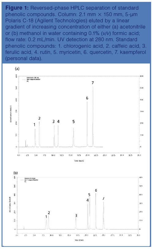
Most of the reversed-phase columns used in either HPLC or UHPLC analysis of phenolic compounds are operated under gradient elution mode with the starting eluent and the gradient former consisting of a water-rich and an organic solvent-rich solution, respectively. A suitable acid is generally incorporated into the starting eluent and, less frequently, into the gradient former solution to control the protonic equilibrium at acidic pH values. Acidic conditions are requested to improve the hydrophobic interactions of the phenolic compounds with the stationary phase by ensuring that both carboxyl and hydroxyl groups of the analytes are in their protonated form. Acetonitrile and methanol are the organic solvents generally used as the gradient former. The two solvents exhibit different elution strength and separation selectivity (Figure 1). However, whenever possible, acetonitrile is preferred to methanol because of its lower UV cut-off and viscosity.

The primary detection method used in LC of phenolic compounds is based on the absorbance of UV or, as in the case of anthocyanins, visible light. These detectors comprise fixed-wavelength, variable-wavelength, scanning, and photodiode array (PDA) detectors. Low-pressure discharge lamps are used as the source of intensive line UV radiations, such as mercury (254 nm) or cadmium (229, 326 nm), whereas deuterium lamps, covering the range 190–700 nm, are used in variable-wavelength and photodiode array detectors, where a tungsten-halogen lamp may also be used to improve the performance in the visible region.
Each class of phenolic compounds has unique spectral characteristics. Therefore, general information on the different classes of phenolic compounds occurring in a complex sample mixture can be obtained by performing UV–vis detection at the wavelength corresponding to the absorption maxima of the phenolic compounds expected to occur in the sample. An example of this approach is depicted in Figure 2, which displays the reversed-phase HPLC separation of phenolic compounds extracted from grape berries and detected by PDA at wavelength values corresponding to the absorption maxima of flavonols (370 nm), anthocyanins (520 nm), and phenolic acids and flavons (320 nm), respectively.

Fluorescence detection is used to detect the limited number of phenolic compounds that naturally fluoresce or that are chemically modified to produce molecules containing a fluorescent tag, usually using on-line postâcolumn derivatization methods. On the other hand, indirect detection, performed by incorporating a fluorescent probe into the mobile phase, as well as chemiluminescence detection with post-column addition of suitable reagents has found limited application.
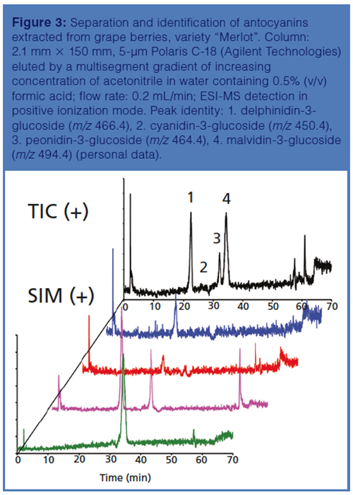
The hyphenation of either HPLC or UHPLC with MS or high resolution mass spectrometry (HRMS), mainly using time-of-flight (TOF) or orbital trap mass analyzers, allows the elucidation of the chemical structure of the investigated phenolic compounds. Prominent among the different ionization sources used in LC–MS is electrospray ionization (ESI) in negative ionization mode, although ESI in positive ionization mode is also used. For example, anthocyanins are glycosylated derivatives of a flavylium cation that are detected by ESI-MS in positive ionization mode (21) (Figure 3). Other, less common, atmospheric pressure ionization (API) interfaces used in LC–MS of phenolics include atmospheric-pressure chemical ionization (APCI) (22) and, to a minor extent, atmospheric-pressure photochemical ionization (APPI) (23).

The hyphenation of liquid chromatography with nuclear magnetic resonance spectroscopy (NMR), which is widely used in analytical chemistry for the unambiguous identification of known and novel organic compounds, is also promising. Direct on-line hyphenation can be realized using flow NMR probes, either of the double saddle Helmholtz coil design or of solenoidal microcoil design, also in combination with methods designed to improve the otherwise low-detection sensitivity of NMR, which is negatively affected by the background absorption of the liquid phase used in the separation step (24). Noticeable among these methods is the hyphenation of LC with NMR using SPE. According to this approach, each phenolic compound eluting the chromatographic column as a separated peak is trapped on an SPE cartridge, which is subsequently dried with nitrogen and then eluted with a deuterated solvent into proper tubes for off-line NMR analysis. An application of this approach has been reported by Goulas et al. for the identification of the methoxylated flavones hispidulin, salvigenin, and cirsimaritin in extracts of Salvia fruticosa, exhibiting antifungal activity (25).
Hydrophilic interaction chromatography (HILIC) is receiving increasing attention, either as a valuable alternative to reversed-phase chromatography or as one of the separation modes used in multidimensional chromatography. The chromatographic retention in HILIC is governed by the hydrophilic partitioning of the analyte between an organic-rich mobile phase and a water layer formed at the surface of a polar stationary phase, with the possible contribution of hydrogen bonding, dipole-dipole interactions, and ion-exchange mechanism. Therefore, HILIC can be considered orthogonal to reversed phase chromatography, whose retention mechanism is based on hydrophobic interactions. On the other hand, similar to reversed-phase chromatography, HILIC uses hydroâorganic mobile phases that are fully compatible with MS detection. Further advantages of HILIC include its suitability for the analysis of polar phenolic compounds that are not sufficiently retained in reversed phase chromatography, and the availability of an alternative separation mechanism, which implies that compounds not easily separated by reversed phase chromatography may be resolved in HILIC. Examples of the applicability of HILIC to the analysis of phenolics include the resolution of individual oligomeric and polymeric procyanidins in apples and apple extracts (26), and the separation and identification of anthocyanins in blueberries, red grape skins, black beans, red cabbage, and red radish (27).
Comprehensive Two-Dimensional Liquid Chromatography
HILIC is very promising for the development of twoâdimensional liquid chromatographic methods for the analysis of phenolic compounds in food and beverages (28). This technique is performed by passing the sample through two columns, each containing a different stationary phase that separate the analytes either according to a diverse separation mechanism or with different selectivity. The technique is performed according to various approaches, which comprise “heart-cutting” and “comprehensive” two-dimensional LC, also referred to as LC-LC and LC×LC, respectively. In LC-LC systems, only the fractions of the effluent from the first column containing the analytes of interest are further separated in the second column, whereas in comprehensive two-dimensional LC the entire effluent from the first column is transferred to the second one.
Comprehensive two-dimensional LC systems, created by coupling HILIC and reversed-phase chromatography, combine the different selectivity and resolving power of the two independent separation mechanisms operating in the first (HILIC) and second (reversed-phase chromatography) columns, which are based on hydrophilicity and hydrophobicity, respectively. HILIC×reversed-phase chromatography techniques are generally operated according to the on-line approach, using a multi-port switching valve with one or two sampling loops, whereas the off-line mode is less popular. On-line, off-line, and stopâflow modalities of HILIC×reversed-phase chromatography have been deeply investigated and successfully applied for the analysis of cocoa procyanidins (29) and for the separation and identification of anthocyanins and their derived pigments in aged red wine (30). Recently, 265 compounds, comprising 196 potentially new phenolic acids, were separated and tentatively characterized in Salvia miltiorrhiza by an off-line two-dimensional HILIC×reversed phase chromatography system, hyphenated to an ion trap time-of-flight mass spectrometer (31). However, because mobile phases with a high content of acetonitrile, such as those used in HILIC, are strong eluents in reversed phase chromatography, system compatibility problems may arise in comprehensive two-dimensional HILIC×reversed-phase chromatography systems.
A less problematic approach of comprehensive twoâdimensional liquid chromatography to the analyses of phenolic compounds is based on the use of two reversedâphase columns of different selectivity. According to this method, phenolic acids and flavonoid antioxidants have been successfully separated in beer and wine using combinations of single or serially coupled polyethylene glycol (PEG)-, phenyl-, and C18- reversed-phase columns in the first dimension and single or two alternating C18 or Zr-carbon columns in the second dimension (32–33). The use of a microbore phenyl column in the first dimension coupled to a monolithic or superficially porous C18 column in the second dimension for comprehensive reversed-phase chromatography×reversed-phase chromatography analysis of phenolic compounds in red wine has also been reported (34). The same group has described a comprehensive twoâdimensional reversed-phase chromatography×reversed-phase chromatography system for the analysis of phenolics in sugarcane leaf extract, consisting of a micro-cyano column and a partially porous C18 column as the first and the second dimension, respectively (35). A total of 34 phenolic compounds, comprising phenolic acids, ellagitannins, flavan-3-ols, flavonols, and ellagic acid conjugates, have been identified in the shoots of Rubus idaeus “Glen Ample” by a twoâdimensional reversed-phase chromatography×reversed-phase chromatography system consisting of a C18 silica column in the first dimension and a pentafluorophenyl column in the second dimension (36).
Besides reversed-phase chromatography and HILIC, other chromatographic separation modes, such as size-exclusion (SEC) or ion exchange chromatography (IEC), have found limited applications in both LC-LC and LC×LC of phenolic compounds. Also limited is the use of dualâretention mechanism columns (37). The combination of SEC with reversed-phase chromatography has been reported for the analysis of green and black teas (Camellia sinensis) (38). The developed comprehensive twoâdimensional SEC×reversed-phase chromatography method has allowed the separation and identification of a variety of phenolic compounds, including catechins, theaflavins and their gallate derivatives, kaempferol, quercetin and myricetin mono-, di-, tri-, and tetraglycosides, esters of quinic acid, and gallic or hydroxycinnamic acids. More recently, a zwitterionic polymethacrylate monolithic column, which provides a dual-retention mechanism, HILIC at high concentrations of acetonitrile, and reversed-phase in water-rich mobile phases, has been used in the first dimension of either HILIC×reversed-phase chromatography or reversed phase chromatography×reversed-phase chromatography comprehensive two-dimensional separations of flavones and related phenolic compounds (39).
Conclusions
To summarize, the expanding interest in phenolic compounds and their positive effects on human health has promoted the development of a variety of techniques for the analysis of these compounds in edible and medicinal plants and in plant-derived food products and dietary supplements. The complexity of such matrices and the heterogeneous chemical structures of the variety of phenolic compounds occurring in the plant kingdom require, besides reliable analytical methods, well-designed sample preparation methods and extraction processes.
Specialized LC separation techniques, combined with state-of-art mass spectrometric detection, are currently used for the separation, identification, and quantification of phenolic compounds in plants and in plant-derived food products. Such techniques comprise HPLC, performed with either analytical size or narrow-bore columns, and UHPLC, with dedicated columns and instrumentation. The majority of HPLC and UHPLC methods currently used for the analysis of phenolic compounds are performed in reversed-phase separation mode, although HILIC is also used, either as a valuable alternative to reversed-phase chromatography or as one of the chromatographic modes used in in 2D LC, in combination with reversed-phase chromatography or with other chromatographic techniques.
References
- B. Wielkopolan and A. ObrÄpalska-StÄplowska, Planta244, 313–332 (2016).
- A.Scalbert, C. Andres-Lacueva, M. Arita, P. Kroon, C. Manach, M. Urpi-Sarda, and D. Wishart, J. Agric. Food Chem.59, 4331–4348 (2011).
- A. Soto-Vaca, A. Gutierrez, J.N. Losso, Z. Xu, and J.W. Finley, J. Agric. Food Chem.60, 6658–6677 (2012).
- U .Lewandowska, K. Szewczyk, E. Hrabec, A. Janecka, and S. Gorlach, J. Agric. Food Chem.61, 12183–12199 (2013).
- M. Iriti and S. Vitalini, Pol. J. Food Nutr. Sci.62, 71–76 (2012).
- J.N. Kabera, E. Semana, A.R. Mussa, and X. He, J. Pharm. Pharmacol.2, 377–392 (2014).
- F. Shahidi and P. Ambigaipalan, J. Funct. Foods18, 820–897 (2015).
- D. Granato, J.S. Santos, L.G. Maciel, and D.S. Nunes, TrAC - Trends Anal. Chem.80, 266–279 (2016).
- E. de Oliveira and R. Battista, Compr. Rev. Food Sci. Food Saf.16, 580–616 (2017).
- V.C. George, G. Dellaire, and H.P.V. Rupasinghe, J. Nutr. Biochem.45, 1–14 (2017).
- M.E. Goetz, S.E. Judd, T.J. Hartman, W. McClellan, A. Anderson, and V. Vaccarino, J. Nutr.146, 2233–2243 (2016).
- A.R. Carvalho, G. Costa, A. Figueirinha, J. Liberal, J.A.V. Prior, M.C. Lopes, M.T. Cruz, and M.T. Batista, Food Res. Int. 99, 485–494 (2017).
- E. Skates, J. Overall, K. De Zego, M. Wilson, D. Esposito, M.A. Lila, and S. Komarnytsky, Food Chem. Toxicol.111, 445–453 (2018).
- L.C. Klein-Júnior, Y. Vander Heyden, and A.T. Henriques, Trends Anal. Chem.80, 66–82 (2016).
- K.-Y. Khaw, M.-O. Parat, P.N. Shaw, and J.R. Falconer, Molecules22, 1286–1208 (2017).
- K. Ameer, H.M. Shahbaz, and J-H. Kwon, Compr. Rev. Food Sci. F. 16, 295–315 (2017).
- S. Tang, H. Zhang, and H.K. Lee, Anal. Chem.88, 228–249 (2016).
- L. Trojer, A. Greiderer, C. Bisjak, W. Wieder, C.W. Huck, and G.K. Bonn, in Handbook of HPLC, D. Corradini, Ed. (CRC Press, Boca Raton, USA, 2nd ed., 2010) pp. 3–45.
- A.A. Rahim, S. Nofrizal, and B. Saad, Food Chem.147, 262–268 (2014).
- P. Chocholouš, J. Vacková, I. Šrámková, D. Šatínský, and P. Solich, Talanta103, 221–227 (2013).
- P. Garcia-Herrera, M.L. Pérez-Rodríguez, T. Aguilera-Delgado, M.J. Labari-Reyes, B. Olmedilla-Alonso, M. Camara, and S. de Pascual-Teresa, Int. J. Food Sci. Technol. 51, 2290–2300 (2016).
- A.P. Singh, T. Wilson, D. Luthria, M.R. Freeman, R.M. Scott, D. Bilenker, S. Shah, S. Somasundaram, and N. Vorsa, Food Chem.127, 80–85 (2011).
- L. Riffault, C. Colas, E. Destandau, L. Pasquier, P. André, and C. Elfakir, Phytochem. Anal. 26, 189–201 (2015).
- R. BrkliaÄa and S. Urban, J. Liq. Chrom. Rel. Technol.34, 1063–1076 (2011).
- V. Exarchou, L. Kanetis, Z. Charalambous, S. Apers, L. Pieters, V. Gekas, and V. Goulas, J. Agric. Food Chem.63, 457–463 (2015).
- W.J. Hollands, S. Voorspoels, G. Jacobs, K. Aaby, A. Meisland, R. Garcia-Villalba, F. Tomas-Barberan, M.K. Piskula, D. Mawson, I. Vovk, P.W. Needs, and P.A. Kroon, J. Chromatogr. A1495, 46–56 (2017).
- C.M. Willemse, M.A. Stander, and A. Villiers, J. Chromatogr. A1319, 127–140 (2013).
- F. Cacciola, S. Farnetti, P. Dugo, P.J. Marriott, and L. Mondello, J. Sep. Sci.40, 7–24 (2017).
- K.M. Kalili and A. de Villiers, J. Chrmatogr. A1289, 69-79 (2013).
- M. Willemse, M. Stander, J. Vestner, A.G.J. Tredoux, and A. de Villiers, Anal. Chem.87, 12006–12015 (2015).
- W. Sun, L. Tong, J. Miao J. Huang, D. Li, Y. Li, H. Xiao, H. Sun, and K. Bi, J. Chromatogr. A1431, 79–88 (2016).
- F. Cacciola, P. Jandera, E. Blahová, and L. Mondello, J. Sep. Sci.29, 2500–2513 (2006).
- E. Blahovà, P. Jandera, F. Cacciola, and L. Mondello, J. Sep. Sci.29, 555–566 (2006).
- P. Dugo, F. Cacciola, M. Herrero, P. Donato, and L. Mondello, J. Sep. Sci.31, 3297–3308 (2008).
- G. Mazzi Leme, F. Cacciola, P. Donato, A.J. Cavalheiro, P. Dugo, and L. Mondello, Anal. Bioanal. Chem.406, 4315–4324 (2014).
- M. Kula D. GÅód, and M. Krauze-Baranowska, J. Pharm. Biomed. Anal.121, 99–106 (2016).
- P. Jandera, M. StaÅková, and T. Hájek, J. Sep. Sci. 36, 2430–2440 (2013).
- C.T. Scoparo, L.M. de Souza, N. Dartora, G.L. Sassaki, P.A.J. Gorin, and M. Iacomini, J. Chromatogr. A1222, 29–37 (2012).
- T. Hájek, P. Jandera, M. StaÅková, and P. Äesla, J. Chromatogr. A1446, 91–102 (2016).
Danilo Corradini earned his Ph.D. in chemistry at Sapienza University of Rome (Italy) and received his postdoctoral education in separation science at the Department of Chemical Engineering, Yale University (USA). His research interests include the study of fundamental and practical aspects of HPLC and electromigration techniques for analytical separations of biomolecules, mainly of plant origin. In recognition of his scientific contributions, he has received the Csaba Horváth Memorial Award in 2009, the Central European Group for Separation Science Award in 2011 and the Arnaldo Liberti Medal in 2014.
Francesca Orsini is a registered nutritionist in the register of the Italian Biologists. She received her bachelor degree in food science and human nutrition in 2012 and master’s degree in food technology and science nutrition in 2014. She completed her education with a Ph.D. in bioengineering and biosciences in 2018 at the University Campus Bio-Medico of Rome. Her research domains include development of sample preparation procedures and chromatographic analytical methods for bioactive compounds in plant material.
Laura De Gara is Full Professor of plant physiology and head of the master’s degree in food science and human nutrition at the Campus Bio-Medico University of Rome. She has almost 30 years of experience in studying plant metabolism with particular attention to redox homeostasis and signalling in response to environmental conditions. She is also interested in plant metabolites with nutritional and healthy effects, in particular polyphenols, fructans, and other fibres, as well as antioxidant vitamins.
Isabella Nicoletti was educated as a biologist at Sapienza University of Rome, where she was also a postdoctoral fellow at the Department of Chemistry from 1982 to 1985. Since 1985 she has been a research scientist at the Institute of Chemical Methodologies of the Italian Research Council in Montelibretti (Rome). She is an expert in the field of separation science, and is interested in the development of chromatographic techniques for innovative analytical methods for foods with determined healthy prerogatives.

AOAC International Awarded NIST Grant for Developing Drug Testing Standards
October 31st 2024The grant will be part of a new collaborative scientific initiative to address the need for standards that define the desired performance of lateral flow immunoassay test strips to detect illicit drugs in tablets and powders.
AI and GenAI Applications to Help Optimize Purification and Yield of Antibodies From Plasma
October 31st 2024Deriving antibodies from plasma products involves several steps, typically starting from the collection of plasma and ending with the purification of the desired antibodies. These are: plasma collection; plasma pooling; fractionation; antibody purification; concentration and formulation; quality control; and packaging and storage. This process results in a purified antibody product that can be used for therapeutic purposes, diagnostic tests, or research. Each step is critical to ensure the safety, efficacy, and quality of the final product. Applications of AI/GenAI in many of these steps can significantly help in the optimization of purification and yield of the desired antibodies. Some specific use-cases are: selecting and optimizing plasma units for optimized plasma pooling; GenAI solution for enterprise search on internal knowledge portal; analysing and optimizing production batch profitability, inventory, yields; monitoring production batch key performance indicators for outlier identification; monitoring production equipment to predict maintenance events; and reducing quality control laboratory testing turnaround time.